Required section · Section 4 of 6
Working the guided case
The guided case specimen is a clean-catch urine received 35 minutes after collection, mixed, and prepared by a validated sediment procedure, examined by bright-field microscopy at 400x with one phase-contrast image in the set. No preservative is declared. The reagent-strip panel shows specific gravity 1.008, pH 8.0 at the upper boundary of the local reference interval, blood 2+, leukocyte esterase trace, and nitrite negative.
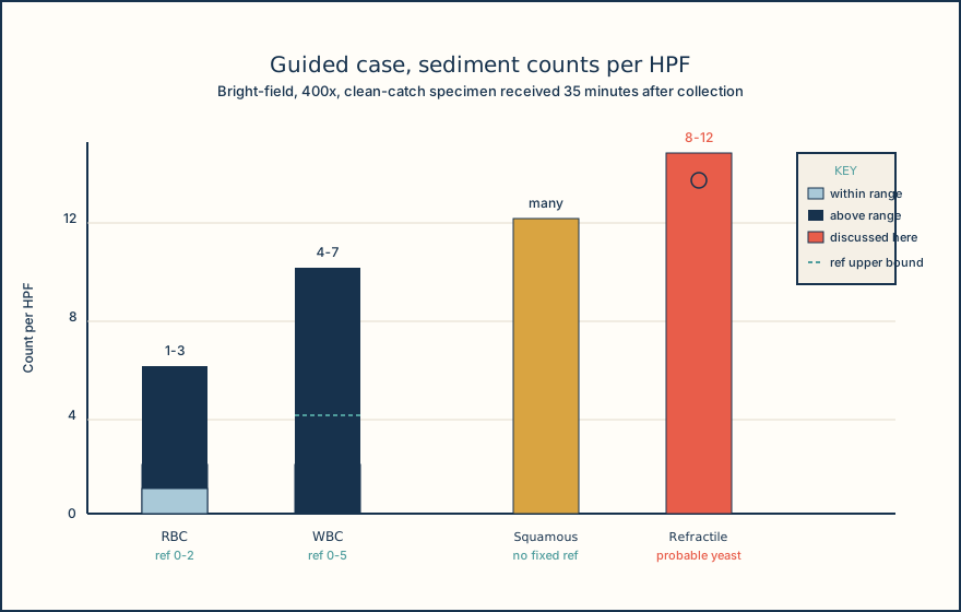
On microscopy, RBCs are 1-3/HPF against a local reference interval of 0-2/HPF, but many appear as pale rings rather than intact discs. The low specific gravity and upper-boundary alkaline pH favor RBC lysis, and the pale rings are compatible with partially lysed RBCs; the strip blood result supports blood-related heme activity in the specimen but does not prove that every ring seen under the microscope is an intact red cell. WBCs are 4-7/HPF against a reference interval of 0-5/HPF, a modest increase, and the trace leukocyte esterase provides a correlating but not confirmatory signal, since the strip reflects enzyme activity rather than a direct cell count.
Squamous epithelial cells are present in large numbers, which weakens confidence that the specimen reflects a well-collected midstream sample and prompts a note on collection quality rather than a change to the interpretation of the other findings. The small round refractile structures, 8-12/HPF against no expected baseline, show repeatable oval walls on repeated viewing, and two of them show narrow-based budding visible in a separate focus plane; that combination of a defined wall and budding is stronger evidence for yeast than the structures' size alone, which is close to that of an RBC.
Putting the findings together: the pale RBC rings, the mild WBC elevation with trace LE, the many squamous cells, and the probable yeast form a set of correlated but individually limited observations. None of them, alone or together, establishes a urinary tract infection, a fungal infection, or a specific diagnosis; microscopy is a morphological and semiquantitative examination, not a method that determines organism identity, viability, or antimicrobial susceptibility. Release the validated observations as classified, note the collection-quality concern from the squamous cell count, and follow the local review or culture-referral policy for the probable yeast and elevated counts rather than naming a diagnosis.
Illustrative drawing — this picture was drawn rather than captured.
| Result | Value | Local reference interval | Flag |
|---|---|---|---|
| Specific gravity | 1.008 | 1.005-1.030 | none |
| pH | 8.0 | 5.0-8.0 | upper boundary |
| Reagent-strip blood | 2+ | negative | positive |
| Leukocyte esterase | trace | negative | positive |
| Nitrite | negative | negative | none |
| RBCs | 1-3/HPF, many pale rings | 0-2/HPF | high |
| WBCs | 4-7/HPF | 0-5/HPF | high |
| Squamous epithelial cells | many/HPF | local method-dependent | review collection quality |
| Small round refractile structures | 8-12/HPF | none expected | review, probable yeast |
Knowledge checks
Reading and checks are open. Sign in only to save.
Knowledge check 1
Knowledge check 2
Section status
Finish this section
Reading and checks are open. Sign in only to save.
The module finishes after every required section is marked done and every check in those sections is correct.