Some problems cannot be resolved with routine testing, such as panreactive plasma, IgG-coated cells that cannot be typed, or a mixture of antibodies. Special methods address a defined laboratory question by changing the reagent, red cells, or antibody mixture. Interpretation depends on what the method detects, which reactivity it removes, which antigens it alters, and which control confirms that the procedure performed as expected.
Antihuman globulin reagents
Antihuman globulin (AHG) binds the human IgG and complement fragments named on the reagent label. The direct antiglobulin test (DAT) detects coating acquired by red cells in the circulation. The indirect antiglobulin test (IAT) produces sensitization during incubation in the laboratory, then detects the bound globulin after the applicable washing or separation step.1,2
Scroll sideways to see all columns
| AHG reagent | Main laboratory use | Interpretation limit |
|---|---|---|
| Monospecific anti-IgG | Detects IgG coating in a DAT or IAT procedure | Reactivity follows the product's stated IgG specificity and any light-chain activity |
| Monospecific anti-C3d | Defines complement coating in a DAT procedure | A reactive result identifies the detected C3 fragment; it does not establish the antibody specificity or cause of coating |
| Polyspecific AHG | Detects IgG plus the complement components named on the label, commonly C3b/C3d or C3d | The exact anti-complement and anti-immunoglobulin coverage is product-specific |
AHG products may use polyclonal, monoclonal, or blended antibodies. The antibody source affects epitope coverage and product performance, so choose a reagent by its label and instructions for use. An anti-C3d reagent has no anti-C4 activity unless the product separately states that coverage. Interpret a reactive DAT or IAT with hemolysis evidence, transfusion and pregnancy history, drug exposure, and the reaction pattern.1,2
Tube-test controls and failure points
In a conventional tube IAT, incubation permits antibody uptake, saline washes remove unbound protein, AHG is added promptly, and calibrated centrifugation produces the endpoint. A negative result is valid only when the procedure's IgG-sensitized control cells agglutinate after their addition. Complement-detecting methods also use appropriate complement-sensitized controls at the frequency specified by the procedure. If a required control fails, repeat the test. Agglutination of the control cells confirms that the AHG remained active. Each product supplies its own cell concentration, serum or plasma volume, incubation, wash, centrifugation, reading, and daily quality control requirements.2,3
For example, the Alba Bioscience tube anti-IgG reagent uses 2 drops of serum or plasma with 1 drop of a 2% to 4% red-cell suspension, incubates at 37 ± 1 °C for 30 to 60 minutes, washes 3 to 4 times, and then centrifuges the AHG test at 900 to 1,000 × g for approximately 10 seconds. These values apply only to that product. A local procedure follows the instructions for the reagent in use.3
Scroll sideways to see all columns
| Failure point | Possible result | Required response |
|---|---|---|
| Residual unbound globulin after inadequate washing | Free AHG is neutralized, producing a false-negative reaction | Repeat the test with the complete validated wash sequence |
| Delay after the final wash | Bound antibody may elute before AHG is added | Add AHG within the procedure's stated interval |
| Contaminated or unsuitable saline | Nonspecific agglutination or loss of cell-bound antibody | Replace the saline and repeat the affected testing |
| Incorrect centrifugation or resuspension | Weak reactions may be dispersed, or compacted cells may resemble agglutination | Verify the calibrated setting and repeat according to procedure |
| Nonreactive sensitized control cells after a negative tube test | The negative result is invalid | Repeat the test from the required step |
The Blood Group Immunology lesson explains IgG sensitization, enhancement media, reaction phases, and complement. The pretransfusion testing process places these methods within antibody investigation and unit selection.
Enzymes, enhancement media, and lectins
Proteolytic enzymes such as ficin and papain alter red-cell membrane proteins and can strengthen, weaken, or remove antigen expression. Compare an enzyme-treated panel with untreated cells from the same investigation. A reaction that disappears suggests an antibody directed against an enzyme-sensitive antigen. A stronger reaction can reveal an enzyme-enhanced specificity. Exclude antibodies to antigens the enzyme weakens or destroys with the untreated panel, because the treated cells have lost those antigens.1,4
Scroll sideways to see all columns
| Typical effect of ficin or papain | Blood group examples | Interpretation consequence |
|---|---|---|
| Enhanced | Rh, Kidd, Lewis, P1, and I | Stronger reactivity can separate one specificity from a mixture |
| Destroyed or markedly weakened | Duffy, M, and N | Treated cells cannot exclude antibodies to the affected antigens |
| Variable | S, s, and other method-sensitive antigens | Use the specific enzyme, concentration, exposure, and reagent-cell instructions |
Enzyme effects vary with the reagent and procedure. An enzyme panel is a supplemental method with its own controls. Albumin, low-ionic-strength solution (LISS), and polyethylene glycol (PEG) change the sensitization environment without enzymatically altering antigen expression. Use them as specified by the validated antibody-detection system.1,4
Lectins bind selected carbohydrate structures. Dolichos biflorus anti-A1 and Ulex europaeus anti-H support ABO subgroup investigations under their reagent instructions. Interpret their patterns with forward and reverse grouping, reaction strength, and the patient's history.1
Adsorption and elution
When a broadly reactive antibody reacts with every panel cell, adsorption can remove it so that an underlying alloantibody can be detected. Adsorption removes selected antibody from serum or plasma by binding it to cells or another antigen-bearing material. Test the adsorbed specimen for reactivity that the removed antibody had masked.
Scroll sideways to see all columns
| Adsorption approach | Best use | Main limit |
|---|---|---|
| Autologous adsorption | Removes autoantibody with the patient's own red cells so residual alloantibody can be investigated | Circulating donor red cells after recent transfusion can adsorb a true alloantibody; severe anemia may leave too few patient cells |
| Allogeneic adsorption | Removes broad autoantibody when recent transfusion or inadequate patient cells prevents autologous adsorption | Selected adsorbing cells can remove alloantibody; the cell set must preserve the ability to detect clinically significant specificities |
| Specialized adsorbent | Removes a defined interference, such as selected cold autoantibody or HLA-related reactivity | Each adsorbent has additional targets and prohibited downstream uses |
Rabbit erythrocyte stroma (RESt) can adsorb selected cold autoantibodies. It can also bind anti-B and anti-P1. Do not use RESt-treated serum for reverse grouping or crossmatching. Repeated adsorption can dilute a weak alloantibody, so the final interpretation records the adsorption performed and the remaining exclusions. The warm AIHA testing pathway explains when autoantibody removal is used in a patient investigation.1,5
Elution releases antibody from coated red cells. The recovered fluid is the eluate. Method selection depends on the suspected specificity, available cell volume, required antibody recovery, and whether the cells must remain suitable for further testing.1,4
Scroll sideways to see all columns
| Elution approach | Common application | Cell and antibody limit |
|---|---|---|
| Acid elution | Recovery of warm-reactive non-ABO IgG | Recovery varies by specificity; some acid methods alter selected antigens |
| Heat at approximately 56 °C | Investigation of ABO antibody coating | Heat damages the red cells and gives poor recovery for many non-ABO antibodies |
| Lui freeze-thaw | ABO hemolytic disease of the fetus and newborn (HDFN) investigation when only a small red-cell volume is available | A total-elution method with poor recovery for many non-ABO antibodies |
| Gentle heat, chloroquine, or another partial-elution procedure | Removal of coating while retaining cells for selected studies | Antibody removal and antigen preservation depend on the procedure |
Organic-solvent elution methods are now uncommon because they lyse the red cells, expose staff to hazardous chemicals, and offer no routine advantage over safer validated methods.1
Wash the sensitized cells until the final wash supernatant is nonreactive, then perform the elution. Test that last wash in parallel with the eluate. A reactive last wash indicates residual free antibody and invalidates the eluate interpretation.4
For a validated ABO HDFN elution, test the eluate with A1, B, and O cells and include the last-wash control. The O cell helps identify broad or unrelated reactivity. The HDFN laboratory pathway explains neonatal interpretation, including the limited role of routine elution.
Antibody titration
Titration estimates relative antibody concentration through twofold serial dilutions. The reported titer is the reciprocal of the highest dilution that meets the laboratory's defined reaction endpoint. An optional score assigns values to every reaction grade and adds them across the dilution series, which can reveal a change spread across several tubes or wells.1,4
Serial comparison requires the same validated method, endpoint, reagent-cell phenotype, cell source, and test conditions. Test the stored baseline specimen beside the current specimen when possible. Prepare dilutions in the direction specified by the procedure and use calibrated pipettes to limit carryover and volume error. Tube, column, and solid-phase titers can produce different numeric endpoints, so the report includes the method used.4
One commonly used score assigns 12 to 4+, 10 to 3+, 8 to 2+, and 5 to 1+. Within that scoring system, a change of 10 or more is commonly treated as significant. The procedure must name the adopted score and its validated cutoff. Separating passive from immune anti-D requires Rh immune globulin history, timing, the reaction pattern, and the trend. The HDFN titer section connects the laboratory result with fetal-risk assessment.1
Mixed red-cell populations and molecular testing
Recent transfusion can place donor and patient red cells in the same specimen. Serologic antigen typing then reflects both populations. Reference laboratories may enrich a patient-cell fraction. Each method has limits on how completely it separates the populations.
Scroll sideways to see all columns
| Separation method | Principle | Main limit |
|---|---|---|
| Differential agglutination | Antiserum agglutinates one population so another fraction can be recovered | Requires a known antigen difference and can leave residual cells from the unwanted population |
| Density or microhematocrit separation | Younger, less dense patient reticulocytes concentrate apart from older donor cells | Depends on adequate reticulocytosis and validated collection of the enriched layer |
| Hypotonic lysis | One reference-laboratory method exposes suitable Hb S-containing specimens to approximately 0.3% saline, then washes the surviving fraction until gross hemolysis subsides | Exact conditions require local validation; enrichment is incomplete and limited to suitable hemoglobinopathy specimens |
Test a nonseparated specimen in parallel when the procedure requires a mixed-field control. Record the separation method and its residual donor-cell risk with the phenotype.
Blood-group genotyping uses DNA from nucleated cells or another validated specimen to predict an antigen profile. Targeted PCR with sequence-specific primers, probe hybridization, arrays, and DNA sequencing can identify defined alleles or nucleotide changes. Genotyping is useful after recent transfusion, when cells are strongly DAT-positive, or when typing reagents are limited. It also supports extended donor typing and rare-donor searches.6,7
Genotyping predicts antigen expression. Serology demonstrates the antigen expressed on the tested cells. Assay panels cover defined alleles and variants. A result can remain uncertain when a variant falls outside that coverage, when a variant changes expression in an unexpected way, or when hematopoietic transplantation and chimerism make the specimen source unrepresentative of the patient's inherited genotype. Use current ISBT allele terminology and report the method's coverage and limitations.6-8
Testing cell-free DNA in maternal plasma can predict selected fetal red-cell antigens within the assay's validated gestational-age and population claims. The HDFN lesson explains the clinical decision that follows.
Donor nucleic acid testing
Donor nucleic acid testing (NAT) detects pathogen DNA or RNA before serologic markers become detectable. Licensed assays use platform-specific PCR or transcription-mediated amplification (TMA). FDA-authorized products define the specimen, analytes, pool size, controls, and intended use. Current examples include multiplex HIV-1/HIV-2, HCV, and HBV assays and separate West Nile virus (WNV) assays. Window-period estimates depend on the assay, pooling, specimen, infection stage, and study design, so interpret any estimate for the specific assay in use and current FDA testing policy.9
Neutralization and thiol reagents
Neutralization uses soluble antigen to bind one antibody in a serum or plasma mixture. Divide the specimen into a neutralized aliquot and a dilution-control aliquot. The control receives an equal volume of saline or the control material named by the procedure. Loss of reactivity only in the neutralized aliquot supports inhibition of the targeted antibody. Investigate any remaining reactivity for clinically significant alloantibodies.1,4
Scroll sideways to see all columns
| Targeted antibody | Validated soluble material may include | Interpretation warning |
|---|---|---|
| Anti-P1 | Commercial P1 substance or another validated P1-bearing preparation | Historical biological sources have variable concentration and safety |
| Anti-Lea or anti-Leb | Soluble Lewis substance | The dilution control distinguishes inhibition from simple dilution |
| Anti-Chido or anti-Rodgers | Complement-containing pooled serum or plasma | The neutralized specimen requires complete alloantibody exclusion |
| Anti-Sda | Validated Sda-bearing material | Use follows reference-laboratory procedure and source controls |
| Anti-I | Validated I-bearing material | Human milk is a historical reference-laboratory source that requires source-safety, validation, and dilution controls |
High-titer, low-avidity (HTLA) describes a serologic pattern of weak, broad AHG reactivity against high-prevalence antigens. Classical examples include antibodies in the Chido/Rodgers, Knops, and JMH systems. These antibodies usually have little effect on red-cell survival, but their broad reactivity can mask a clinically significant alloantibody. Neutralization, selected enzyme effects, thiol treatment, rare cells, and reference-laboratory testing help define the interference.1
Dithiothreitol (DTT) and 2-mercaptoethanol reduce disulfide bonds and can disperse IgM pentamers in a treated serum aliquot. Compare treated and untreated aliquots under one validated method when the laboratory question is whether IgM or IgG accounts for reactivity. The result depends on reagent concentration, exposure time, temperature, and controls. When the dilution control remains reactive, a known IgM control loses activity, and an IgG control retains activity, loss of the patient's reactivity supports an IgM contribution. A nonreactive treated aliquot shows only that no IgG reactivity was detected. Weak IgG may remain below the method's detection limit. The reagent red cells remain untreated in this serum-treatment comparison.10
DTT treatment of reagent red cells has a different purpose. It removes anti-CD38 therapeutic interference by destroying CD38 on the cells, and it also destroys Kell and weakens or destroys other DTT-sensitive antigens. AABB's updated 2026 guidance recommends K-negative units after DTT-based testing unless the patient is known to be K-positive. Antibodies to k, Yta, Dombrock, and other DTT-sensitive antigens may not be detected. Baseline or current genotyping, the antibody history, DTT-treated controls, and the approved unit-selection process help address this residual risk.11
Column and solid-phase endpoints
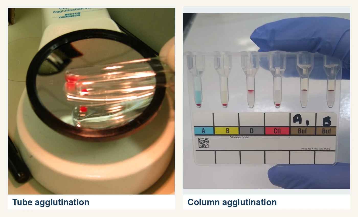
Tube, column-agglutination, and solid-phase methods create different visible endpoints. The reagent card, plate, instrument, and instructions for use define the analyte, controls, cell concentration, incubation, centrifugation, wash sequence, and grading rules.12,13
Column agglutination
In a column card, plasma or serum and reagent cells incubate in the reaction chamber above the gel. Centrifugation then moves unagglutinated cells through the porous matrix to a pellet at the bottom. Agglutinates remain near the top or disperse through the column according to their size. A mixed-field result contains an agglutinated population in the column and an unagglutinated pellet at the bottom. Reaction grades and matrix composition are product-specific.13
Scroll sideways to see all columns
| Illustrative column pattern | Appearance |
|---|---|
| 4+ | Dense band of agglutinates at the top |
| 3+ | Most agglutinates in the upper half |
| 2+ | Agglutinates distributed through the column |
| 1+ | Agglutinates concentrated in the lower half, with some cells at the bottom |
| Negative | Compact cell pellet at the bottom |
| Mixed field | Agglutinated cells in the column plus a pellet at the bottom |
Read the card within the product's stated interval. Fibrin, particulates, rouleaux, incorrect cell concentration, drying, and improper centrifugation can create anomalous patterns. Patient transfusion and transplantation history helps determine whether a mixed-field reaction represents two real cell populations. Correlate the pattern with card quality and specimen findings. Column methods incorporate separation within the matrix and use their own positive and negative quality controls.13
Solid phase
Solid-phase formats bind red-cell membrane, antigen, antibody, or Protein A to a microplate well. In one FDA-authorized Protein A format, patient serum or plasma and reagent red cells incubate in the coated well. Washing removes unbound protein, anti-IgG links sensitized cells, and centrifugation produces a smooth cell layer for a positive reaction. Unsensitized cells form a compact central button. Other systems use red-cell membrane-coated wells and indicator cells, so the endpoint and controls are specific to that assay.12
Each solid-phase system has defined indications for antibody screening, identification, crossmatch, DAT, or antigen typing. The product label defines the analyte. Platelet and HLA enzyme-linked or multiplex assays use related surface-bound principles with their own antigen panels and signal systems. Interpret the assay through its named analyte, controls, and intended use.
Chloroquine and EGA treatment
An IgG-positive DAT can interfere with antigen typing that uses an IAT phase. Chloroquine diphosphate and EDTA-glycine-acid (EGA) are partial-elution treatments that can remove cell-bound IgG and leave the cells suitable for selected antigen studies. Their antigen effects differ.1,4
Scroll sideways to see all columns
| Treatment | Useful effect | Principal limit |
|---|---|---|
| Chloroquine diphosphate | Dissociates IgG and preserves many red-cell antigens | Can leave complement on the cell; some saline-reactive monoclonal antisera, including Rh typing reagents, are unsuitable after treatment |
| EGA | Rapidly dissociates IgG under a validated procedure | Denatures Kell-system antigens and makes treated cells unsuitable for K/k assignment |
Confirm IgG removal with monospecific anti-IgG after chloroquine treatment because complement can remain and react with polyspecific AHG. Test a known antigen-positive cell through the same treatment when the method requires it. Exposure time, temperature, reagent concentration, and overtreatment limits belong in the procedure. After recent transfusion, the treated cells remain unreliable for assigning the patient's inherited phenotype, even after the coating antibody is removed. Use genotyping or reference-laboratory testing to resolve that problem. For every method, record what it removed or altered so that later reviewers know which antibodies it could not exclude.1,5
Practice
References
- Bloch EM, Campbell-Lee S, McKenna DH Jr, Montemayor-Garcia C, Schwartz J, Shaz B, Storry J, eds. Technical Manual. 22nd ed. AABB; 2026. Back to text
- Diagast. Anti-Human Globulin Anti-IgG, Anti-C3d, and Anti-IgG,-C3d Instructions for Use. US Food and Drug Administration. Accessed August 29, 2026. Back to text
- Alba Bioscience Limited. Anti-Human Globulin Anti-IgG Instructions for Use. US Food and Drug Administration. Accessed August 29, 2026. Back to text
- Raman L, Armstrong B, Smart E. Principles of laboratory techniques. ISBT Sci Ser. 2020;15(suppl 1):81-111. doi:10.1111/voxs.12591 Back to text
- Johnson ST, Puca KE. Evaluating patients with autoimmune hemolytic anemia in the transfusion service and immunohematology reference laboratory: pretransfusion testing challenges and best transfusion-management strategies. Hematology Am Soc Hematol Educ Program. 2022;2022(1):96-104. doi:10.1182/hematology.2022000406 Back to text
- Association for the Advancement of Blood & Biotherapies. Standards for Molecular Testing for Red Cell, Platelet, and Neutrophil Antigens. 7th ed. AABB; 2024. Effective January 1, 2025. Back to text
- US Food and Drug Administration. Establishment of Reference Panels for Blood Group Genotyping Tests. Accessed August 29, 2026. Back to text
- International Society of Blood Transfusion. ISBT Blood Group Database. August 2026 release. Accessed August 29, 2026. Back to text
- US Food and Drug Administration. Complete List of Donor Screening Assays for Infectious Agents and HIV Diagnostic Assays. Accessed August 29, 2026. Back to text
- Okuno T, Kondelis N. Evaluation of dithiothreitol (DTT) for inactivation of IgM antibodies. J Clin Pathol. 1978;31(12):1152-1155. doi:10.1136/jcp.31.12.1152 Back to text
- Association for the Advancement of Blood & Biotherapies. Association Bulletin #16-02: Mitigating the Anti-CD38 Interference With Serologic Testing. Revised March 2026. Back to text
- Bio-Rad Medical Diagnostics GmbH. Solidscreen II Microplate for Solid Phase Antiglobulin Tests With TANGO Instruments Instructions for Use. US Food and Drug Administration. Accessed August 29, 2026. Back to text
- Diagnostic Grifols, SA. DG Gel 8 Cards Instructions for Use. US Food and Drug Administration. Accessed August 29, 2026. Back to text