Required section · Section 2 of 6
Read zones as shared migration neighborhoods
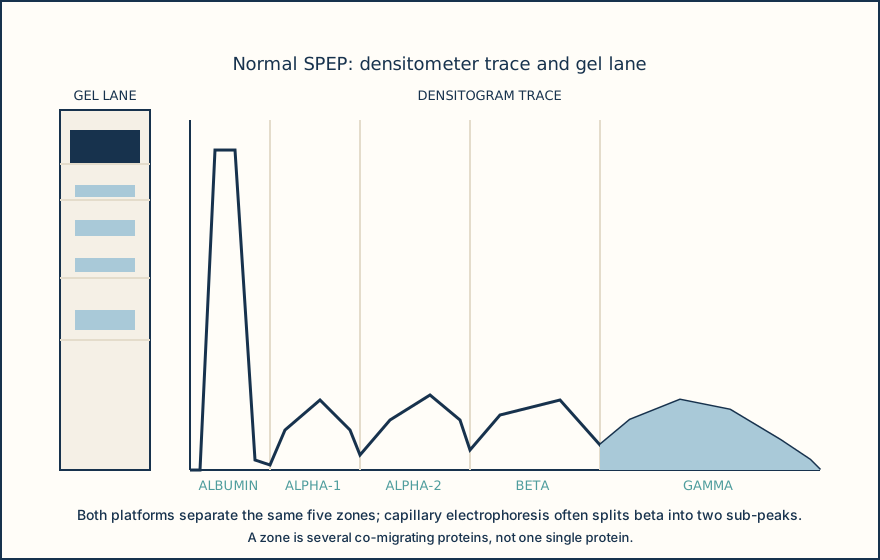
SPEP separates serum proteins by charge and size into albumin, alpha-1, alpha-2, beta, and gamma zones. These zones are migration neighborhoods, not single purified proteins. Albumin is normally the dominant band and largest fraction.
The gamma zone is dominated by immunoglobulins. A broad diffuse gamma rise is a polyclonal pattern, while a narrow sharply bordered peak is a restricted feature that needs characterization. Beta and gamma can overlap, so location alone is not enough.
Use the trace shape and the paired gel lane to label fractions before attaching meaning. Describe width and border before calling a pattern restricted or polyclonal.
Illustrative drawing — this picture was drawn rather than captured.
Reusable first-pass reading sequence for a serum trace.
Verify the specimen
Confirm properly clotted serum and review hemolysis or collection concerns.
Label the zones
Locate albumin, alpha-1, alpha-2, beta, and gamma before interpreting shape.
Describe the pattern
Record broadness, border, distribution, and beta-gamma continuity.
Select follow-up
Use specimen review and local protocol to repeat, perform IFE, or correlate sFLC.
Knowledge checks
Reading and checks are open. Sign in only to save.
Knowledge check 1
Section status
Finish this section
Reading and checks are open. Sign in only to save.
The module finishes after every required section is marked done and every check in those sections is correct.