Required section · Section 1 of 6
A restricted gamma pattern arrives at the bench
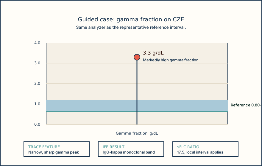
A serum protein electrophoresis (SPEP) trace from a 68-year-old patient shows total protein 8.6 g/dL, above the local reference interval of 6.4-8.3 g/dL. The gamma fraction is 3.3 g/dL, and the trace has a narrow, sharply bordered peak. Whether this is a true restricted protein pattern and what study should follow depends on specimen and interference review.
SPEP separates proteins into visible migration zones, then estimates fraction concentration from the trace area. It is a screening and characterization result, not an immunoglobulin type or disease diagnosis. A narrow peak can be meaningful, but it also needs specimen and interference review.
Start with what the instrument shows: zone, width, border, and specimen status. Release only the supported pattern description and follow the local reflex pathway.
Illustrative drawing — this picture was drawn rather than captured.
| Analyte | Result | Reference interval | Trace observation |
|---|---|---|---|
| Total protein | 8.6 g/dL | 6.4-8.3 g/dL | Delta check: prior 6.9 g/dL |
| Albumin | 3.4 g/dL | 4.02-4.76 g/dL | Low |
| Gamma | 3.3 g/dL | 0.80-1.35 g/dL | Narrow, sharply bordered peak |
Knowledge checks
Reading and checks are open. Sign in only to save.
Knowledge check 1
Section status
Finish this section
Reading and checks are open. Sign in only to save.
The module finishes after every required section is marked done and every check in those sections is correct.