A high potassium result on a specimen with red-tinged serum, with acceptable quality control on the analyzer, calls for finding where in the testing cycle the error began. Patient physiology, specimen collection and handling, the measurement system, and result reporting all affect a laboratory result. Locating a discrepancy within the testing cycle directs the investigation toward the records and corrective actions most likely to explain it.1,2
The testing cycle
The terms preanalytic, analytic, and postanalytic describe three connected phases. Current standards also use preexamination, examination, and postexamination for the same sequence.
Scroll sideways to see all columns
| Phase | Boundary | Representative failures | First evidence to review |
|---|---|---|---|
| Preanalytic | Test selection and ordering through specimen arrival and preparation for analysis | Wrong patient or test, incomplete preparation, wrong container, short draw, clot, contamination, hemolysis, delayed transport, or unsuitable storage | Order, patient identifiers, collection record, container, specimen appearance, transport conditions, and receipt time |
| Analytic | The measurement procedure | Reagent deterioration, calibration error, failed quality control, instrument malfunction, pipetting error, or method-specific interference | Quality-control and calibration records, reagent lot and expiration, maintenance, flags, interference indices, and instrument messages |
| Postanalytic | Result verification through delivery to the ordering clinician | Transcription or interface error, unsuitable reference interval, missed critical-result notification, uninvestigated delta, or incorrect corrected report | Result history, reference interval, autoverification rules, notification record, corrected-report record, and audit trail |
A defect can cross phase boundaries. Prolonged contact with cells begins as a handling problem and changes the specimen before analysis. Hemolysis begins during collection or transport and produces an analyte-dependent effect during measurement. The investigation follows the event from its origin through the reported result.
Patient preparation and biologic variation
Patient state can change a measured concentration even when the collection and method perform correctly. Name any preparation that affects interpretation in the order and collection instructions.3,4
Scroll sideways to see all columns
| Variable | Laboratory consequence | Control |
|---|---|---|
| Time of day | Cortisol, iron, therapeutic drugs, and other measurands can vary with circadian rhythm or dosing time. | Collect at the defined time and record it when interpretation depends on timing. |
| Food and fasting | Meals can change glucose and triglycerides and can make the specimen lipemic. | Apply the test-specific fasting instruction and document preparation. |
| Posture and tourniquet use | Standing and prolonged venous stasis concentrate proteins, protein-bound substances, and cells. Fist pumping and prolonged stasis can change several measurands. | Standardize posture when required, allow about 15 minutes of rest after a recent posture change, avoid fist pumping, and release the tourniquet as blood flow begins. |
| Exercise and stress | Recent exertion or acute stress can change lactate, creatine kinase, leukocytes, catecholamines, and related results. | Record relevant exertion or stress and delay elective collection when the procedure specifies a rest period. |
| Medication and supplement use | A drug may change the patient's physiology, react in the measurement procedure, or create a recognizable assay artifact. | Record relevant exposure and use the current assay instructions during investigation. |
| Peak and trough timing | Drug concentration changes across the dosing interval. | Collect a trough immediately before the next dose. The drug and the laboratory procedure set when to collect a peak. |
Posture changes and prolonged bed rest have different time courses. A brief change to a recumbent position shifts fluid into the circulation. Prolonged head-down bed-rest studies have observed reduced plasma volume and hemoconcentration. Collection posture and the duration of immobilization therefore matter when comparing serial results.4,5
Intravenous crystalloid administration can lower hemoglobin, hematocrit, and albumin concentrations. A volunteer study found an albumin fall greater than dilution alone explained, consistent with redistribution as well. Infusion fluid that enters the collection tube directly contaminates the specimen, which is a separate problem.6
Age affects the appropriate comparison interval. Alkaline phosphatase, for example, can rise during childhood and adolescent bone growth and requires age-appropriate interpretation.7 Current cigarette smoking is associated with higher leukocyte counts. Longitudinal observations show a decrease after cessation.8 Preparation and exposure history help interpret a result alongside the reporting laboratory's validated interval.
CMS interprets 42 CFR §493.1291(d) to require the intervals supplied to clients to reflect the method and, when applicable, the patient population.3
Hormone therapy can shift some sex-associated laboratory results. Current Association for Diagnostics & Laboratory Medicine (ADLM) guidance recommends analyte-specific interpretation that considers the hormone regimen and duration, organ inventory, muscle mass, and the laboratory's validated reporting policy. After at least 6 months of stable gender-affirming hormone therapy, hemoglobin, hematocrit, and red-cell results generally follow the interval associated with the affirmed gender. Creatinine and estimated glomerular filtration rate require additional care because muscle mass and the sex input used by the calculation can alter the estimate, and ADLM reports no consensus for that input. Dual calculation or cystatin C may help when kidney assessment remains uncertain.9
Specimen integrity and interference
An interference creates a clinically significant difference in a result because another substance or property of the specimen affects the measurement procedure. The direction and size of the effect depend on the analyte, concentration of the interferent, specimen matrix, reagent, analyzer, and calculation used. A normal serum or plasma specimen is clear and pale yellow. Hemolysis, icterus, and lipemia each change that appearance. Check the specimen at receipt and confirm with the analyzer indices. Because the effect depends on the method, a visible specimen change cannot support one correction factor across methods.2,10
Scroll sideways to see all columns
| Finding | Main mechanisms | Laboratory response |
|---|---|---|
| Hemolysis | Red-cell contents enter plasma or serum; free hemoglobin adds spectral and chemical effects; intracellular fluid can dilute extracellular constituents. Potassium, lactate dehydrogenase (LD), aspartate aminotransferase (AST), phosphate, and other measurands may change. | Use the analyzer's hemolysis index and the laboratory's analyte-specific interference limits. Suppress, qualify, repeat, or request recollection according to the validated policy and clinical urgency. |
| Icterus | Bilirubin absorbs light and can participate in chemical reactions. | Apply the method's icterus index and manufacturer interference claim. Use a validated blank, wavelength correction, alternate method, or recollection pathway when appropriate. |
| Lipemia | Turbidity scatters light; a reduced plasma-water fraction can affect methods that dilute the sample. | Apply the method's lipemia index. A validated ultracentrifugation, dilution, blank, or alternate method may resolve the effect. |
| Clot or microclot | Inadequate mixing or unsuitable anticoagulation allows fibrin formation. | Reject or process only under a test-specific validated procedure. Inspect analyzer flags and probes when a clot is discovered after aspiration. |
| Infusion contamination | Fluid or medication from a vascular line dilutes or adds constituents to the specimen. | Review the collection site and infusion, then recollect from an acceptable site or follow the validated line-draw procedure. |
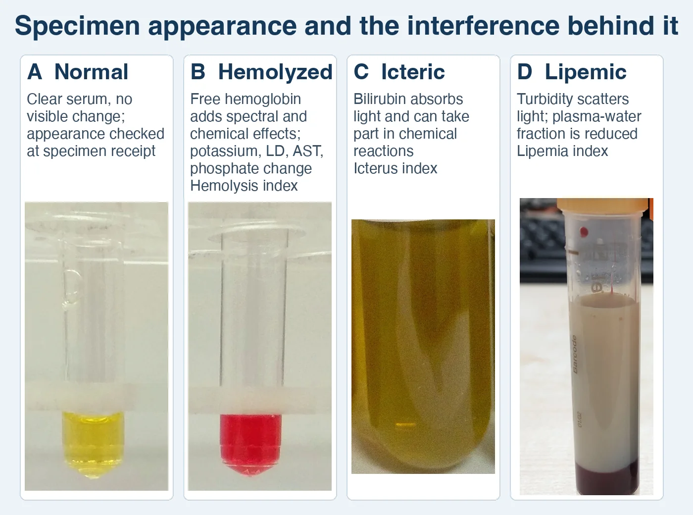
Collection-related hemolysis can follow a needle or device that produces excessive shear, forceful syringe aspiration or transfer, vigorous mixing, collection before antiseptic dries, or unsuitable transport. Match the device to the vein and required flow, allow antiseptic to dry, fill tubes through the closed collection system when available, mix by gentle inversion, and use validated transport conditions.4
Pseudohyperkalemia and pseudohypokalemia. In vitro red-cell injury, fragile leukemic cells, extreme thrombocytosis, or delayed separation can raise measured potassium without a matching increase in the patient's circulating plasma. Marked metabolically active leukocytosis can consume potassium after collection and lower the measured result. Compare serum and plasma when appropriate, review the blood count and collection history, and repeat promptly with a properly collected and handled specimen. The size of the artifact cannot be predicted reliably from visual hemolysis alone.11
Pseudohyponatremia. Severe hyperlipidemia or hyperproteinemia reduces the water fraction of plasma. An indirect ion-selective electrode dilutes the specimen and can report a falsely low sodium concentration under these conditions. A direct ion-selective electrode measures undiluted specimen and generally avoids this electrolyte-exclusion artifact. Measured osmolality and the clinical setting help identify the discrepancy.12
Assay-specific examples
Scroll sideways to see all columns
| Interferent | Pattern | Investigation |
|---|---|---|
| Excess biotin | Some streptavidin-biotin sandwich immunoassays produce falsely low results; some competitive assays produce falsely high results. Direction and magnitude follow the assay design. | Review supplements, dose, renal function, collection time, package insert, and laboratory policy. Use a validated alternate method or recollect after the assay-specific interval. |
| Heterophile or human anti-animal antibodies | Antibody bridging or blocking can produce a falsely high or low immunoassay result. | Compare with the clinical pattern and prior results. Serial dilution, blocking reagent, an alternate platform, or reference-laboratory testing may help during the investigation. |
| Anti-CD38 therapy | Daratumumab and isatuximab can cause panreactivity during indirect antiglobulin testing and can mask an alloantibody. | Notify the transfusion service and follow its Blood Banking procedure. |
| Emicizumab | Activated partial thromboplastin time-based factor VIII and inhibitor assays can give misleading results during therapy, and the effect may persist for up to 6 months after the last dose. | Use assay classes identified as suitable in the current prescribing information and the coagulation laboratory's procedure. |
| Nucleic-acid amplification inhibitors | Heparin, heme compounds, and other matrix constituents can inhibit amplification. Poor stabilization can degrade the target, and amplified product can contaminate later reactions. | Follow the test-specific collection and stabilization instructions. Keep preamplification and postamplification work separate and use the validated contamination-control workflow. |
The FDA describes clinically important biotin interference and recommends that device developers characterize and communicate it. Take any biotin withholding interval from the current assay instructions. A single universal interval cannot account for dose, kidney function, assay tolerance, and measurement design.13 A current AABB association bulletin covers anti-CD38 interference in transfusion testing, and the current emicizumab prescribing information identifies affected coagulation assays.14,15
Ordering, identification, and acceptability
Patient and specimen identification remain linked from the order through the final report. Use at least two person-specific identifiers and label the specimen container in the patient's presence. Never use a room number or physical location as an identifier.1,16
CLIA permits an oral test request when the laboratory solicits written or electronic authorization within 30 days and documents its effort to obtain it. A laboratory or institution may set a stricter local policy. The requisition and specimen must supply enough information to identify the patient, authorized requester, requested examination, specimen source when relevant, and collection date and time when those details affect testing.17
The laboratory or institution defines the turnaround expected for stat and as soon as possible requests. The collection and receipt times show whether the specimen met that local definition.
Written acceptability criteria list each defect, its consequence, and the action:
Scroll sideways to see all columns
| Finding | Main concern | Action |
|---|---|---|
| Unlabeled specimen or identifier mismatch | Result can be assigned to the wrong patient. | Reject under the identification policy. Apply the documented irreplaceable-specimen exception only when the policy permits it and the responsible parties resolve identity. |
| Wrong container or additive | The method lacks the required matrix or receives a chemical interferent. | Reject or seek documented approval under a validated exception. |
| Underfilled citrate tube | Excess citrate relative to plasma can prolong clotting results. | Reject according to the coagulation procedure.18-20 |
| Hematocrit above 55% in a citrate tube | The reduced plasma volume leaves excess citrate relative to plasma, prolonging clotting results even in a correctly filled tube. | Adjust the volume of citrate in the tube using the collection-device instructions and the laboratory's validated high-hematocrit calculation. The citrate reagent itself stays at full strength.18-20 |
| Clot in anticoagulated blood | Cell counts fall and analyzer probes may obstruct; coagulation testing becomes unsuitable. | Reject the affected testing. |
| Hemolysis, icterus, or lipemia | The effect varies by analyte and method. | Apply the analyte-specific interference policy. |
| Delay or temperature excursion | Cells, enzymes, organisms, or measurands may change before analysis. | Compare the documented time and temperature with the test's validated stability limit. |
| Leaking container | Specimen integrity and worker safety are compromised. | Contain the leak, follow the exposure procedure when needed, and request recollection under policy. |
Blood collection tubes and order of draw
Whether a tube suits a test depends on its additive and the manufacturer's instructions. Stopper colors can vary. Current CLSI venous-collection guidance uses this sequence to limit additive carryover: blood culture, sodium citrate, serum, heparin, EDTA, then glycolytic inhibitor.21,22
Scroll sideways to see all columns
| Sequence | Additive or specimen | Function and common use |
|---|---|---|
| 1 | Blood-culture medium | Supports recovery of microorganisms; collection technique protects volume and sterility. |
| 2 | 3.2% buffered sodium citrate | Reversibly binds calcium. A 9:1 blood-to-anticoagulant ratio supports routine coagulation testing; fill the tube to the manufacturer-specified volume.18-20 |
| 3 | Serum, with or without clot activator and separator gel | Produces serum after clotting. Gel and clotting requirements come from the test and tube instructions. |
| 4 | Lithium or sodium heparin | Potentiates antithrombin and limits clot formation. Commonly used for plasma chemistry. |
| 5 | K2EDTA | Chelates calcium and preserves cellular morphology. Commonly used for hematology and selected molecular tests. |
| 6 | Glycolytic inhibitor | Fluoride slows glycolysis. Prompt separation or another validated stabilizing system may still be required for glucose. |
Fill additive tubes to the required volume and mix them by the tube manufacturer's gentle inversion schedule. Shaking can damage cells. Delayed or inadequate mixing can allow microclots. EDTA carryover can produce a markedly increased potassium with a markedly decreased calcium. Separator-gel effects on drugs and other measurands depend on tube composition, contact time, and method.
Venous collection follows the laboratory's procedure: confirm the order and preparation, identify the patient, select and clean the site, allow antiseptic to dry, collect in the required sequence, release the tourniquet as flow begins, mix additive tubes, label at the bedside, and document the collector and time. Fist pumping and tourniquet use longer than one minute increase preanalytic variation.4
Special collections require their own validated instructions. A line draw controls infusion contamination and dead space. An arterial blood-gas specimen controls air exposure, anticoagulant, mixing, temperature, and time to analysis. Capillary collection controls puncture site and depth, warming, first-drop handling, and tissue-fluid contamination. How cerebrospinal fluid is divided among tubes and transported depends on the tests requested and the local tube-routing policy. Urine timing, preservation, and acceptability are covered in Specimen Handling and Acceptability and Urine Specimen Types.
Analytic and postanalytic safeguards
The analytic phase begins only after the specimen has met acceptance criteria. Reagent identity, preparation, storage, and expiration; calibration status; quality-control results; maintenance; instrument flags; and validated interference rules must support release. Under the general CLIA minimum for quantitative testing, the laboratory tests at least two control materials of different concentrations each day it performs patient testing. An individualized quality control plan, a specialty requirement, or a more stringent manufacturer instruction can replace that default with its own design. Run controls also after the reagent, calibration, maintenance, and critical-part events the procedure specifies, and release patient results only after the control results are acceptable.17
Postanalytic review checks whether the result fits the specimen, method, patient history, reference interval, and laboratory release rules. A delta check compares the current value with a prior value using analyte- and patient-specific thresholds established by the laboratory. Autoverification applies validated rules for flags, quality control, range, interference, and consistency before releasing a result without manual review.
Critical-result communication follows the laboratory's written policy. Under 42 CFR §493.1291(g), the laboratory immediately alerts the person or entity requesting the test and, when applicable, the person responsible for using the result when a result indicates an imminently life-threatening condition or a panic or alert value. CMS interpretive guidance says the record should include the date, time, result, and person notified.3,17
Corrected reports follow a separate requirement. Under 42 CFR §493.1291(k), when the laboratory detects an error in a reported patient result, it promptly notifies the authorized person who ordered the test and, when applicable, the person using the result; promptly issues a corrected report; and maintains both the original and corrected reports. How long records are kept depends on the document type and on any longer state or accreditation requirement.3,17
Pretest probability and result interpretation
Sensitivity and specificity describe performance against a reference classification. Pretest probability describes the estimated probability of the condition before the new result. Bayes reasoning combines these quantities to estimate probability after testing.23
Probability of disease after a positive result = (sensitivity × pretest probability) ÷ [(sensitivity × pretest probability) + ((1 − specificity) × (1 − pretest probability))]
Probability of no disease after a negative result = (specificity × (1 − pretest probability)) ÷ [(specificity × (1 − pretest probability)) + ((1 − sensitivity) × pretest probability)]
- After a positive result: (0.92 × 0.10) ÷ [(0.92 × 0.10) + (0.04 × 0.90)] = 0.092 ÷ 0.128 = 71.9% probability of disease.
- After a negative result: (0.96 × 0.90) ÷ [(0.96 × 0.90) + (0.08 × 0.10)] = 0.864 ÷ 0.872 = 99.1% probability of no disease.
The same test result gives a different posttest probability when the pretest probability changes. Before you apply a performance claim, check the population, setting, patient selection, specimen quality, and reference classification behind it.
Practice
References
- Clinical and Laboratory Standards Institute. Patient and Laboratory Specimen Identification Processes. 1st ed. CLSI standard PRE01. Clinical and Laboratory Standards Institute; 2024. Accessed August 28, 2026. https://clsi.org/shop/standards/pre01/ Back to text
- Clinical and Laboratory Standards Institute. Handling, Transport, Processing, and Storage of Blood Specimens for Routine Laboratory Examinations. 1st ed. CLSI guideline PRE04. Clinical and Laboratory Standards Institute; 2023. Accessed August 28, 2026. https://clsi.org/shop/standards/pre04/ Back to text
- Centers for Medicare & Medicaid Services. State Operations Manual, Appendix C: Survey Procedures and Interpretive Guidelines for Laboratories and Laboratory Services. Updated January 23, 2026. Accessed August 29, 2026. Back to text
- Simundic AM, Bölenius K, Cadamuro J, et al. Joint EFLM-COLABIOCLI recommendation for venous blood sampling. Clin Chem Lab Med. 2018;56(12):2015-2038. doi:10.1515/cclm-2018-0602 Back to text
- Trudel G, Uhthoff HK, Laneuville O. Hemolysis during and after 21 days of head-down-tilt bed rest. Physiol Rep. 2017;5(24):e13469. doi:10.14814/phy2.13469 Back to text
- Lobo DN, Stanga Z, Simpson JA, Anderson JA, Rowlands BJ, Allison SP. Dilution and redistribution effects of rapid 2-litre infusions of 0.9% (w/v) saline and 5% (w/v) dextrose on haematological parameters and serum biochemistry in normal subjects: a double-blind crossover study. Clin Sci (Lond). 2001;101(2):173-179. Back to text
- Mayo Clinic Laboratories. Alkaline phosphatase, serum. Pediatric Catalog. Test ID ALP. Accessed September 7, 2026. Back to text
- Higuchi T, Omata F, Tsuchihashi K, Higashioka K, Koyamada R, Okada S. Current cigarette smoking is a reversible cause of elevated white blood cell count: cross-sectional and longitudinal studies. Prev Med Rep. 2016;4:417-422. doi:10.1016/j.pmedr.2016.08.009 Back to text
- Association for Diagnostics & Laboratory Medicine. ADLM guidance document on incorporating gender diversity in pathology and laboratory medicine. Published July 2026. Accessed August 28, 2026. Back to text
- Clinical and Laboratory Standards Institute. Interference Testing in Clinical Chemistry. 3rd ed. CLSI guideline EP07 Plus. Clinical and Laboratory Standards Institute; 2018. Reaffirmed October 2022. Accessed August 28, 2026. https://clsi.org/shop/standards/ep07-plus/ Back to text
- Simundic AM, Baird G, Cadamuro J, Costelloe SJ, Lippi G. Managing hemolyzed samples in clinical laboratories. Crit Rev Clin Lab Sci. 2020;57(1):1-21. doi:10.1080/10408363.2019.1664391 Back to text
- Aziz F, Sam R, Lew SQ, Massie L. Pseudohyponatremia: mechanism, diagnosis, clinical associations and management. J Clin Med. 2023;12(12):4076. doi:10.3390/jcm12124076 Back to text
- US Food and Drug Administration. Testing for Biotin Interference in In Vitro Diagnostic Devices: Guidance for Industry. October 2020. Accessed August 28, 2026. Back to text
- Association for the Advancement of Blood & Biotherapies. Mitigating the Anti-CD38 Interference with Serologic Testing. Association Bulletin #16-02. Revised March 2026. Accessed August 28, 2026. Back to text
- Genentech, Inc. Hemlibra (emicizumab-kxwh) prescribing information. Revised July 2025. Accessed August 28, 2026. Back to text
- The Joint Commission. Two patient identifiers: understanding the requirements. Updated April 21, 2026. Accessed August 28, 2026. Back to text
- Centers for Medicare & Medicaid Services. 42 CFR §§493.1105, 493.1241, 493.1242, 493.1256, and 493.1291. Electronic Code of Federal Regulations. Updated August 27, 2026. Accessed August 28, 2026. Back to text
- Clinical and Laboratory Standards Institute. Collection, Transport, and Processing of Blood Specimens for Testing Plasma-Based Coagulation Assays. 6th ed. CLSI standard H21. Clinical and Laboratory Standards Institute; 2024. Accessed August 29, 2026. https://clsi.org/shop/standards/h21/ Back to text
- Becton, Dickinson and Company. BD Vacutainer Evacuated Blood Collection System instructions for use. US catalog locator, English document VDP40384, revision 09, March 28, 2026. Accessed September 8, 2026. Back to text
- Siemens Healthineers Academy. Hemostasis: basic overview online training. Accessed August 29, 2026. Back to text
- Clinical and Laboratory Standards Institute. Order of blood draw tubes and additives. Published March 19, 2019. Accessed August 29, 2026. Back to text
- Clinical and Laboratory Standards Institute. Collection of Diagnostic Venous Blood Specimens. 8th ed. CLSI standard PRE02. Clinical and Laboratory Standards Institute; 2025. Back to text
- Altman DG, Bland JM. Diagnostic tests 1: sensitivity and specificity. BMJ. 1994;308:1552. doi:10.1136/bmj.308.6943.1552 Back to text