When a leukocyte count or differential falls outside the reference interval, the first step is to decide whether the change is reactive. Evaluate it with the absolute counts, the differential, cell maturity, and blood-film morphology. Marrow release, redistribution between circulating and marginated pools, altered cell survival, infection, inflammation, medication effects, and specimen aging can all change the result. Persistent unexplained findings, blasts, or a uniform abnormal population call for targeted testing for a clonal disorder.1,2
Absolute counts and reference intervals
A relative differential divides a fixed total among the leukocyte lines. An absolute count shows the measured concentration of each line and prevents a change in one population from being mistaken for a change in another. The ASCP BOC examination reference ranges page contains the ASCP BOC composite values for examination review. Interpret patient results against your laboratory's verified interval for the patient's age and population.3,4
The teaching thresholds below describe a pattern. Interpret patient results against the local interval:
Scroll sideways to see all columns
| Cell line | Increased absolute count | Decreased absolute count |
|---|---|---|
| Neutrophils | Adult neutrophilia is often taught as an absolute neutrophil count (ANC) above about 7.0 × 10³/µL. Demargination produces a rapid mature neutrophilia; infection, inflammation, tissue injury, corticosteroids, and growth-factor exposure are other causes. | Severe neutropenia is an ANC below 0.5 × 10³/µL. Infection risk depends on depth, duration, mucosal integrity, marrow reserve, and the clinical setting. Prolonged severe neutropenia carries the greatest risk. |
| Eosinophils | An absolute eosinophil count of at least 0.5 × 10³/µL is a common definition of eosinophilia. Persistent counts of at least 1.5 × 10³/µL meet the quantitative definition of hypereosinophilia. Helminth infection, atopy, drug reactions, and clonal disorders are major categories. | A low or zero result has little meaning in isolation. Corticosteroids and acute stress can lower the count. |
| Basophils | Persistent basophilia can accompany hypersensitivity, chronic inflammation, and myeloid neoplasia. The magnitude, duration, other CBC findings, and film determine the next test. | A low count usually has little independent diagnostic value. |
| Monocytes | Monocytosis occurs during recovery from marrow suppression or acute infection and with chronic infection, inflammatory disease, tissue injury, and neoplasia. Persistent unexplained monocytosis requires evaluation for a clonal process. | Monocytopenia can accompany marrow suppression, corticosteroid exposure, severe infection, or hemodialysis. |
| Lymphocytes | Adult lymphocytosis is an absolute lymphocyte count above the laboratory's adult upper reference limit; teaching thresholds commonly fall around 4–5 × 10³/µL.18 Pediatric upper limits are higher and change rapidly through infancy and childhood. | Adult lymphocytopenia is commonly taught as a count below 1.0 × 10³/µL. Pediatric lower limits are age-specific. Infection, immune deficiency, systemic disease, and cytotoxic or immunosuppressive treatment are common settings. |
Current consensus definitions support the severe-neutropenia and eosinophilia thresholds shown here.5,6
Age-partitioned pediatric intervals are especially important during the neonatal period and early childhood. A single child cutoff can misclassify a normal developmental count.4
The Duffy-null erythrocyte phenotype caused by ACKR1 variation is associated with a lower baseline ANC without increased infection risk. Race is an unreliable substitute for phenotype or genotype. A 2026 multinational study found a central 95% adult ANC interval of 1.21 to 5.39 × 10³/µL in its United States Duffy-null cohort, with variation among study sites. These values support phenotype-aware intervals. Each laboratory establishes its own interval. A falling count, symptoms, other cytopenias, or discordant clinical findings still require investigation.7,8
Left shift and marked reactive leukocytosis
A left shift is an increase in immature neutrophil-lineage cells in blood, usually bands and sometimes metamyelocytes, myelocytes, or promyelocytes. It can occur with neutrophilia, a normal ANC, or neutropenia. Report immature stages by name. CLSI H20 separates bands and segmented neutrophils in its reference visual differential and recognizes that some instruments combine them. Document which cells your laboratory's ANC includes, so serial results stay comparable.2,9
Tell the stages apart by their granules and nuclear shape. Myeloblasts and promyelocytes have large, round nuclei, and azurophilic primary granules appear at the promyelocyte stage. Specific secondary granules are made in myelocytes and metamyelocytes, as the round nucleus becomes kidney-shaped. The nucleus then takes a curved band shape before a segmented neutrophil forms.19
Suppose a CBC shows a WBC count of 10.0 × 10³/µL, 55% segmented neutrophils, 4% bands, 2% metamyelocytes, and 1% myelocytes. Under an ANC definition that includes segmented neutrophils and bands:
ANC = (segmented neutrophil fraction + band fraction) × WBC = (0.55 + 0.04) × 10.0 × 103/µL = 5.9 × 10³/µL
Report the metamyelocytes and myelocytes separately, by name, so the left shift stays visible.
A leukemoid reaction is marked reactive neutrophilic leukocytosis, traditionally a WBC count above 50 × 103/µL with a left shift. This threshold is an operational convention. Severe infection or inflammation, metabolic stress, growth-factor exposure, and cytokine-secreting tumors can produce the pattern. The blood film and clinical setting guide testing for chronic myeloid leukemia (CML) and other myeloid neoplasms.1,10
Scroll sideways to see all columns
| Finding | Leukemoid reaction pattern | CML pattern |
|---|---|---|
| Neutrophil morphology | Toxic granulation, Döhle bodies, and vacuoles may accompany a left shift | A broad granulocytic maturation spectrum is typical; dysplastic forms can occur |
| Basophils and eosinophils | Usually lack the persistent combined increase typical of CML | Basophilia is characteristic; eosinophils can also increase |
| Hemoglobin and platelets | Results follow the underlying illness and can be normal or abnormal | Anemia and thrombocytosis or abnormal platelet morphology can support the pattern |
| Leukocyte alkaline phosphatase (LAP) | Usually increased | Often decreased in untreated disease |
| Defining study | Reactive cause established and clonal testing selected when indicated | BCR::ABL1 fusion demonstrated by a validated molecular or cytogenetic method |
CML diagnosis requires a BCR::ABL1 result interpreted in the full clinicopathologic setting.11
LAP scoring is now mainly of historical interest. One hundred bands and segmented neutrophils are graded from 0 to 4+, and the score is the sum of each grade multiplied by the number of cells at that grade. A count with 18 cells at 4+, 28 at 3+, 32 at 2+, 14 at 1+, and 8 at 0 gives:
LAP score = (18 × 4) + (28 × 3) + (32 × 2) + (14 × 1) + (8 × 0) = 234
Interpret any score with your laboratory's own interval. LAP can support a reactive explanation for the leukocytosis. Evaluating CML still requires the molecular or cytogenetic studies described above.10,11,12
A leukoerythroblastic blood film contains immature myeloid cells together with nucleated red cells, and that combination defines the pattern. Anemia, neutrophilia, and teardrop cells may accompany it, and prominent teardrop cells strongly support marrow fibrosis or infiltration. Marrow infiltration or fibrosis, severe infection, shock or trauma, brisk hemolysis, and recovery from marrow suppression are recognized settings. The hypoproliferative anemia lesson explains the myelophthisic pattern in marrow replacement.13
Reactive neutrophil morphology
Reactive changes on the film show accelerated production, cell activation, phagocytosis, or degeneration. Each finding is nonspecific, so check that it agrees with the specimen age, CBC, analyzer flags, and clinical setting before you report it.9,14
Scroll sideways to see all columns
| Change | Blood-film appearance | Interpretation |
|---|---|---|
| Toxic granulation | Coarse dark blue-purple cytoplasmic granules | Common with infection, inflammation, tissue injury, and granulocyte colony-stimulating factor exposure |
| Döhle bodies | Pale blue-gray inclusions near the cytoplasmic edge | Aggregates of rough endoplasmic reticulum associated with infection, inflammation, burns, pregnancy, and growth-factor exposure |
| Cytoplasmic vacuoles | Small pinholes or larger clear spaces; organisms may be visible in phagocytic vacuoles | Supports activation or phagocytosis when fresh; prolonged EDTA exposure and alcohol toxicity can produce similar vacuolation. Do not label vacuoles toxic without accompanying toxic changes14 |
| Pyknosis | A single dense, dark, round-to-oval nucleus with homogeneous chromatin | Degenerative change (necrobiosis) that can precede cell death |
| Nuclear fragmentation (karyorrhexis) | Rounded nuclear fragments without filaments or recognizable chromatin pattern | Degenerative change (necrobiosis); when many cells are affected, check the collection time and film-preparation delay |
| Variable neutrophil size | Marked size variation within the same film | Can accompany activated-cell swelling or slide adhesion and has limited value alone |
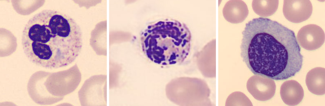
During the first week of ehrlichiosis or anaplasmosis, a film may show purple intracytoplasmic microcolonies called morulae. Anaplasma phagocytophilum favors granulocytes, Ehrlichia chaffeensis favors monocytes, and Ehrlichia ewingii favors granulocytes. Morulae are a rapid presumptive finding with low sensitivity, so a film without them does not exclude infection. The infected cell type does not identify the species. Leukopenia and thrombocytopenia commonly accompany these infections. The vector-borne blood isolate lesson covers specimen choice, PCR, serology, and reporting.15
Reactive lymphocyte patterns
Reactive lymphocytosis commonly accompanies viral infection, including Epstein-Barr virus (EBV), cytomegalovirus, hepatitis viruses, and acute HIV. Pertussis and selected other infections, drug reactions, vaccination, endocrine disease, and acute physiologic stress can also raise the absolute count. Lymphocytopenia occurs with infection, immune deficiency, systemic illness, marrow failure, and immunosuppressive or cytotoxic treatment. Choose the next step from the absolute count, patient age, duration, and film pattern.1,4
ICSH recommends reactive lymphocyte when morphology supports a benign stimulus and abnormal lymphocyte with a description when a clonal process is suspected. The older terms atypical, variant, Downey cell, Türk cell, and immunoblast are historical and inconsistent. Morphology guides follow-up. Flow cytometry or another clonality study establishes clonality.9
Scroll sideways to see all columns
| Population pattern | Typical features | Laboratory action |
|---|---|---|
| Reactive | Heterogeneous cell size, nuclear shape, chromatin density, nucleoli, and cytoplasmic basophilia; abundant cytoplasm can mold around adjacent red cells | Correlate with the clinical setting and targeted infectious or drug testing |
| Persistent uniform abnormal population | Similar cells repeated across the film, sometimes with a disease-associated morphology | Review prior counts and analyzer flags; select flow cytometry or hematopathology review according to laboratory policy |
| Blasts or blast-like cells | High nucleus-to-cytoplasm ratio, fine chromatin, and nucleoli, with variable size and cytoplasm | Confirm urgently and escalate under your laboratory's critical or urgent-review procedure |
A plasmacytoid lymphocyte is a reactive form with eccentric nuclear placement and deeply basophilic cytoplasm. When plasmacytoid forms are numerous or plasma cells circulate, correlate with the clinical setting and consider immunophenotyping, because reactive and clonal processes can share this appearance.
Infectious mononucleosis
EBV infects B lymphocytes through interactions that include viral gp350/220 binding to CD21. The large reactive lymphocytes in infectious mononucleosis are mainly activated EBV-specific CD8-positive T cells responding to infected B cells, with contributions from NK and CD4-positive T cells. The CBC commonly shows absolute lymphocytosis with a heterogeneous reactive population. Mild thrombocytopenia and increased aminotransferases can accompany the pattern.1,16
Scroll sideways to see all columns
| Test or pattern | Interpretation |
|---|---|
| Heterophile antibody | Can support a typical mononucleosis syndrome. False-positive and false-negative results occur, and young children often remain negative. CDC advises against Monospot testing for general use. |
| Viral capsid antigen (VCA) IgM | Appears early and usually disappears within 4 to 6 weeks. VCA IgM with absent EBV nuclear antigen antibody supports primary infection. |
| VCA IgG | Appears during the acute phase, peaks within several weeks, and then persists for life. Read it together with VCA IgM and EBNA antibody. |
| EBV nuclear antigen (EBNA) antibody | Standard immunofluorescence detects it after the acute phase, commonly 2 to 4 months after symptom onset. VCA IgG and EBNA antibodies with absent VCA IgM support past infection. |
The serologic timing, persistence, and pattern interpretations in this table follow CDC guidance.17
More than 90% of adults have evidence of past EBV infection. A high VCA IgG concentration occurs in acute and past infection and does not establish when the infection occurred. Cytomegalovirus and other infections can produce a similar heterophile-negative mononucleosis pattern. Choose and interpret tests according to the clinical presentation and the assay's instructions, and read EBV serology as a pattern across all three markers.17
Practice
References
- Keohane EM, Preston MM, Mirza KM, Walenga JM, eds. Rodak's Hematology: Clinical Principles and Applications. 7th ed. Elsevier; 2025. Accessed August 29, 2026. Back to text
- Clinical and Laboratory Standards Institute. Reference Leukocyte Differential Count (Proportional) and Evaluation of Instrumental Methods. 3rd ed. CLSI standard H20. Published April 29, 2026. Accessed August 29, 2026. Back to text
- Electronic Code of Federal Regulations. 42 CFR §493.1253: Standard: establishment and verification of performance specifications. Accessed August 29, 2026. Back to text
- Bohn MK, Wilson S, Steele S, Adeli K. Comprehensive pediatric reference intervals for 79 hematology markers in the CALIPER cohort of healthy children and adolescents. Int J Lab Hematol. 2023;45(4):469-480. doi:10.1111/ijlh.14068. Back to text
- Fioredda F, Skokowa J, Tamary H, et al. The European guidelines on diagnosis and management of neutropenia in adults and children: a consensus between the European Hematology Association and the EuNet-INNOCHRON COST Action. HemaSphere. 2023;7(4):e872. doi:10.1097/HS9.0000000000000872. Back to text
- Wang SA, Orazi A, Gotlib J, et al. The international consensus classification of eosinophilic disorders and systemic mastocytosis. Am J Hematol. 2023;98(8):1286-1306. doi:10.1002/ajh.26966. Back to text
- Hibbs SP, Chipare I, Halawani AJ, et al. Multinational assessment of absolute neutrophil counts and white blood cell counts among healthy Duffy-null adults. Blood. 2026;147(3):290-298. doi:10.1182/blood.2025029359. Back to text
- Palmblad J, Sohlberg E, Nilsson CC, et al. Clinical and immunological features in ACKR1/DARC-associated neutropenia. Blood Adv. 2024;8(3):571-580. doi:10.1182/bloodadvances.2023010400. Back to text
- Palmer L, Briggs C, McFadden S, et al. ICSH recommendations for the standardization of nomenclature and grading of peripheral blood cell morphological features. Int J Lab Hematol. 2015;37(3):287-303. doi:10.1111/ijlh.12327. Back to text
- Abukhiran IA, Jasser J, Syrbu S. Paraneoplastic leukemoid reactions induced by cytokine-secreting tumours. J Clin Pathol. 2020;73(6):310-313. doi:10.1136/jclinpath-2019-206340. Back to text
- Apperley JF, Milojkovic D, Cross NCP, et al. 2025 European LeukemiaNet recommendations for the management of chronic myeloid leukemia. Leukemia. 2025;39(8):1797-1813. doi:10.1038/s41375-025-02664-w. Back to text
- American Society of Hematology. Leukocyte alkaline phosphatase scoring. ASH Image Bank. Published September 28, 2011. Accessed August 29, 2026. Back to text
- Tabares Calvache E, Tabares Calvache AD, Faulhaber GAM. Systematic review about etiologic association to the leukoerythroblastic reaction. Int J Lab Hematol. 2020;42(5):495-500. doi:10.1111/ijlh.13238. Back to text
- College of American Pathologists. 2026 Hematology, Clinical Microscopy, and Body Fluids Glossary. 2026:3-4. Accessed September 7, 2026. Back to text
- Biggs HM, Behravesh CB, Bradley KK, et al. Diagnosis and management of tickborne rickettsial diseases: Rocky Mountain spotted fever and other spotted fever group rickettsioses, ehrlichioses, and anaplasmosis, United States. MMWR Recomm Rep. 2016;65(RR-2):1-44. doi:10.15585/mmwr.rr6502a1. Back to text
- Damania B, Kenney SC, Raab-Traub N. Epstein-Barr virus: biology and clinical disease. Cell. 2022;185(20):3652-3670. doi:10.1016/j.cell.2022.08.026. Back to text
- Centers for Disease Control and Prevention. Laboratory testing for Epstein-Barr virus (EBV). Updated April 10, 2024. Accessed August 29, 2026. Back to text
- Gloucestershire Hospitals NHS Foundation Trust. Haematology reference ranges. Adult lymphocyte interval. Accessed September 8, 2026. Back to text
- Lawrence SM, Corriden R, Nizet V. The ontogeny of a neutrophil: mechanisms of granulopoiesis and homeostasis. Microbiol Mol Biol Rev. 2018;82(1):e00057-17. doi:10.1128/MMBR.00057-17. Back to text