In the hematology laboratory, a lymphoid neoplasm often first appears as an unexplained lymphocytosis, circulating blasts, or rouleaux that suggest a paraprotein. Lymphoid neoplasms are clonal malignancies of B-, T-, or NK-cell lineage. This group is classified with the same integrated workup used for myeloid disease: CBC and blood film, marrow morphology, flow immunophenotyping, cytogenetics, and molecular testing. Classification here follows the fifth edition of the World Health Organization classification (WHO5). The International Consensus Classification (ICC) differs in selected names and definitions, so state in each report which framework you applied.1,2 The myeloid neoplasia lesson covers the shared background: leukemia and lymphoma patterns, acute and chronic behavior, and the history that moved diagnosis from morphology to genetics. The lymphoid group divides into precursor lymphoblastic neoplasms, mature lymphoid neoplasms, and plasma-cell neoplasms.
Acute lymphoblastic leukemia and lymphoma
Acute lymphoblastic leukemia (ALL) and lymphoblastic lymphoma (LBL) are one disease family. ALL presents with blood and marrow involvement. LBL presents as a tissue mass, often nodal or mediastinal, with limited blood and marrow involvement. B-lineage disease accounts for about 80 to 85% of cases, and ALL remains the most common childhood cancer.3
B-ALL presents with fatigue from anemia, fever from neutropenia-driven infection, and mucocutaneous bleeding from thrombocytopenia. Lymphadenopathy is common, splenomegaly and hepatomegaly can be present, and bone pain reflects intramedullary growth. Central nervous system (CNS) and gonadal involvement can occur, and cerebrospinal fluid (CSF) may contain lymphoblasts when CNS disease is present, so correlate the film and flow findings with any CSF specimen submitted. T-ALL often presents with a mediastinal mass alongside anemia, thrombocytopenia, organomegaly, and bone pain. Teenage males predominate, and circulating blast counts can be high. Leukopenia tends to be less severe in T-ALL than in B-ALL.3
WHO5 classification
WHO5 classifies B-lymphoblastic leukemia/lymphoma (B-ALL) by its defining genetic abnormality. Cases that remain unclassified after a full genetic workup are B-ALL, not otherwise specified (NOS). When testing is limited, the family-level term B-ALL, not further classified, is permitted.1,4
Scroll sideways to see all columns
| WHO5 B-ALL entity | Defining finding | Laboratory note |
|---|---|---|
| B-ALL, NOS | No recurrent abnormality found | Assigned only after full genetic workup |
| With high hyperdiploidy | Modal chromosome number 51 to 67, with a nonrandom gain pattern | Favorable risk; the largest childhood subtype |
| With hypodiploidy | Modal chromosome number 43 or fewer | Adverse risk |
| With iAMP21 | Intrachromosomal amplification of chromosome 21 | About 2% of childhood B-ALL; adverse risk |
| With BCR::ABL1 fusion | t(9;22) Philadelphia chromosome | Adverse risk; adults are disproportionately affected |
| With BCR::ABL1-like features | Kinase-activating lesions producing a BCR::ABL1-like expression profile | Requires transcript-level or sequencing methods |
| With KMT2A rearrangement | KMT2A fusion, most often AFF1 | Common in infant ALL; adverse risk |
| With ETV6::RUNX1 fusion | t(12;21) | Favorable risk; common in childhood |
| With ETV6::RUNX1-like features | Similar expression profile with ETV6 and IKZF1 deletions | New entity in WHO5 |
| With TCF3::PBX1 fusion | t(1;19) | Intermediate risk |
| With IGH::IL3 fusion | t(5;14), driving IL3 overexpression | Associated with eosinophilia |
| With TCF3::HLF fusion | t(17;19) | Rare; adverse risk; newly defined in WHO5 |
| With other defined genetic abnormalities | DUX4, MEF2D, ZNF384, or NUTM1 rearrangement; IGH::MYC; PAX5 alterations or PAX5 p.P80R | Detected mainly by sequencing or transcript methods |
T-ALL keeps two entities in WHO5: T-lymphoblastic leukemia/lymphoma, NOS, and early T-cell precursor (ETP) lymphoblastic leukemia/lymphoma. No genetic subtype is a formal T-ALL entity because the proposed transcription-factor subgroups overlap and lack established clinical relevance. NK-cell lymphoblastic leukemia, a provisional entity in the fourth edition, was removed.4
Morphology and immunophenotype
Lymphoblasts fall into two size classes. The more common small form measures 1.0 to 2.5 times a normal lymphocyte, with scant blue cytoplasm and indistinct nucleoli. The larger form measures 2 to 3 times lymphocyte size, with prominent nucleoli and an irregular nuclear membrane, and it can resemble acute myeloid leukemia (AML) blasts. Morphology establishes that a blast process is present. Lineage and classification come from immunophenotyping and genetics.3
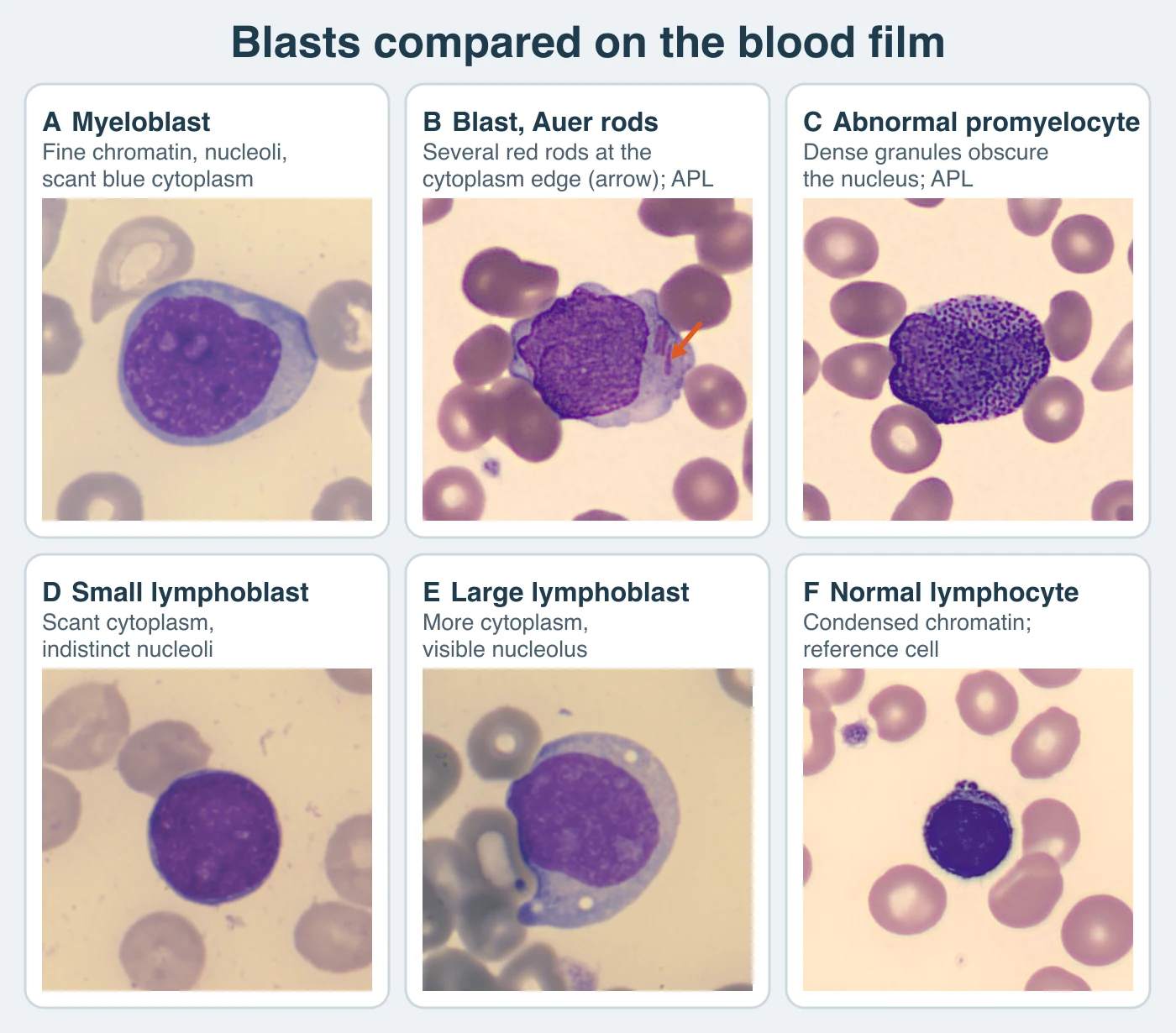
Terminal deoxynucleotidyl transferase (TdT) is common in both B- and T-lineage lymphoblasts. CD34 expression is variable. B-ALL is usually HLA-DR positive. T-ALL is often HLA-DR negative. B-lineage blasts express CD19, CD20, CD22, CD24, CD79a, CD10, cytoplasmic μ heavy chain, and PAX5 in a sequence that tracks maturation. Cytoplasmic or surface CD3 is lineage-defining for T-ALL.3
Scroll sideways to see all columns
| Maturation stage | Immunophenotype | Frequency note |
|---|---|---|
| Early (pro/pre-pre) B-ALL | CD34, CD19, cytoplasmic CD22, TdT | About 5% of childhood ALL and 11% of adult ALL |
| Common (intermediate) B-ALL | CD34, CD19, CD10, cytoplasmic CD22, TdT | Most common childhood subtype |
| Pre-B-ALL | CD34, CD19, cytoplasmic CD22, cytoplasmic μ, variable TdT | Cytoplasmic μ marks the stage |
| T-ALL | Cytoplasmic or surface CD3, CD2, CD4, CD5, CD7, CD8, TdT in most cases; CD34 variable; HLA-DR often negative | Teenage males predominate; mediastinal mass and extramedullary disease are common |
Some immunophenotypes suggest the underlying genetic lesion. Intense CD10 with decreased CD9 and CD20 suggests ETV6::RUNX1. Absent CD10 and CD24 with CD15 or NG2 expression suggests a KMT2A rearrangement. Flow cytometry can therefore suggest a defining lesion before cytogenetic results are available.4
The ETP immunophenotype defines the second T-ALL entity: cytoplasmic CD3 positive, CD7 positive, CD1a negative, CD8 negative, CD5 dim on fewer than 75% of blasts, and coexpression of one or more stem-cell or myeloid markers (CD13, CD33, CD34, CD117, or HLA-DR) on at least 25% of blasts. ETP disease accounts for roughly 5 to 17% of pediatric and about 7% of adult T-ALL. Use a broad panel of myeloid and stem-cell markers so ETP-ALL can be distinguished from acute undifferentiated leukemia and mixed-phenotype disease.4
Genetics and measurable residual disease
Risk assessment integrates age, presenting disease burden, defining genetics, early treatment response, and especially measurable residual disease (MRD). Genetic effects are lesion-specific: high hyperdiploidy and ETV6::RUNX1 are generally favorable in childhood B-ALL. Hypodiploidy, KMT2A-rearranged infant ALL, TCF3::HLF, and iAMP21 are adverse. Flow cytometry or sequencing of immunoglobulin or T-cell receptor gene rearrangements measures MRD, and the result is one of the strongest predictors of relapse in ALL. Other hematology lessons describe the analytic methods.4
Mature lymphoid neoplasms
Mature lymphoid neoplasms arise from lymphoid cells arrested at, or derived from, particular normal stages of B-, T-, or NK-cell development. This developmental-stage model fits some diseases better than others. Hairy cell leukemia, for example, has no clearly identified normal counterpart. Morphology is the first step. Because different entities can look alike on a film, flow cytometry and immunohistochemistry are needed to resolve most cases. Recurrent chromosomal translocations frequently place a growth or survival gene next to an actively transcribed immunoglobulin locus.1,3
Scroll sideways to see all columns
| Entity | Morphology on film or tissue | Immunophenotype and defining genetics | Laboratory recognition |
|---|---|---|---|
| Chronic lymphocytic leukemia (CLL)/small lymphocytic lymphoma | Small mature lymphocytes with dense clumped chromatin and scant cytoplasm; smudge cells common; increased prolymphocytes can be described, and they no longer define a separate CLL/prolymphocytic leukemia entity | CD19 positive, CD20 positive, CD23 positive with aberrant CD5; dim surface immunoglobulin | Diagnostic threshold and prognostic markers in the text below |
| T-cell or NK large granular lymphocytic leukemia | Abundant pale-blue cytoplasm with medium-large azurophilic granules | T-LGL: CD3, CD8, CD57 positive with clonal T-cell receptor (TCR) rearrangement. NK-LGL: surface CD3 negative, CD16 positive, with a restricted killer-cell immunoglobulin-like receptor (KIR) pattern providing supportive evidence of clonality when integrated with persistence, phenotype, and molecular findings | Physiologic LGL counts reach about 15% of lymphocytes or 0.6 × 10³/µL; leukemia is classically above 2 × 10³/µL, though lower clonal counts with compatible findings occur.5 Aggressive NK-cell leukemia is a separate entity |
| Hairy cell leukemia | Round or ovoid nuclei without prominent nucleoli and abundant cytoplasm with circumferential hairy projections | Classic phenotype CD19, CD20, CD22, CD11c, CD25, CD103, CD123 positive; BRAF V600E in most cases;6 annexin A1 supports the diagnosis | Marrow fibrosis often produces a dry tap, so a trephine biopsy is important. WHO5 renames the biologically distinct former hairy cell leukemia variant as splenic B-cell lymphoma/leukemia with prominent nucleoli (SBLPN) |
| T-prolymphocytic leukemia | Medium-sized cells with one prominent nucleolus and cytoplasmic blebs; small-cell and cerebriform variants occur | Mature pan-T phenotype, usually CD2, CD3, CD5, CD7 positive; TCL1A rearrangement common | Aggressive course; WHO5 no longer recognizes B-cell prolymphocytic leukemia as a distinct entity, so such cases are reclassified among mature B-cell neoplasms |
| Follicular lymphoma | Circulating cells show condensed chromatin with distinct nuclear clefts; marrow biopsy shows a paratrabecular tumor distribution | t(14;18) IGH::BCL2 or, less often, BCL6 rearrangement | Blood involvement occurs in about 10% of patients |
| Mantle cell lymphoma | Small-cell form can mimic CLL or marginal zone lymphoma; blastoid variant can mimic ALL or diffuse large B-cell lymphoma (DLBCL) | t(11;14) IGH::CCND1 with cyclin D1 overexpression in most cases; cyclin-D1-negative disease requires SOX11 or CCND2/CCND3 evidence | An indolent variant is restricted to blood, marrow, and spleen |
| Burkitt lymphoma/leukemia | Medium-sized cells with finely clumped chromatin and deeply basophilic, vacuolated cytoplasm; biopsy shows a starry-sky pattern from tingible-body macrophages | Mature B cell with surface immunoglobulin, light-chain restriction, and CD10; TdT and CD34 negative | Endemic, sporadic, and immunodeficiency-associated epidemiologic variants;7 WHO5 recommends distinguishing EBV-positive and EBV-negative Burkitt lymphoma; IG::MYC translocation; WHO5 recognizes the separate entity high-grade B-cell lymphoma with 11q aberrations.1 Marrow and CNS involvement affect staging, and the high tumor burden makes tumor lysis syndrome a laboratory risk (see the myeloid neoplasia lesson) |
| Diffuse large B-cell lymphoma and high-grade B-cell lymphoma | Diffuse proliferation of large B cells | Integrated morphology, phenotype, and genetics are required; WHO5 separates DLBCL/HGBL with dual MYC and BCL2 rearrangements. MYC and BCL6 double-rearranged disease is classified by morphology | Marrow involvement may occur and is often focal |
| Mycosis fungoides and Sézary syndrome | Cerebriform or folded nuclei with scant cytoplasm; Sézary cells can be large or small in the same patient; skin biopsy may show Pautrier microabscesses in a minority | Effector-memory phenotype in mycosis fungoides; central-memory phenotype in Sézary syndrome | Blood criteria in the text below |
| Anaplastic large cell lymphoma | Large pleomorphic hallmark cells; marrow disease can be subtle | Strong uniform CD30; ALK rearrangements define ALK-positive ALCL | Primary cutaneous and breast-implant-associated ALCL are distinct entities from systemic ALK-negative ALCL |
| Peripheral T-cell lymphoma, NOS | Heterogeneous; defined by exclusion | T-cell antigen mismatch, most often absent CD7 or CD5 | Marrow involvement in 20 to 30%; eosinophilia can accompany the disease |
Chronic lymphocytic leukemia
CLL is the most common adult leukemia in Western populations, with a median age of about 72. Many patients are asymptomatic and found through a routine CBC with absolute lymphocytosis of small mature lymphocytes and smudge cells. The International Workshop on CLL diagnostic threshold is at least 5 × 10³/µL clonal B cells in blood sustained for at least 3 months. A lower count without lymphadenopathy, organomegaly, disease-related cytopenias, or symptoms is monoclonal B-cell lymphocytosis. A cytopenia caused by a typical marrow infiltrate can establish CLL despite a lower blood B-cell count. Flow cytometry demonstrates clonality by showing the characteristic phenotype: CD19, CD20 (dim), CD23, and aberrant CD5 with dim surface immunoglobulin and light-chain restriction. CLL with unmutated IGHV arises from a pre-germinal-center cell and has an aggressive course. CLL with mutated IGHV arises from a post-germinal-center cell and has an indolent course, making IGHV mutation status a major prognostic factor. TP53 disruption is a major predictive variable.8
Hodgkin lymphoma
WHO5 retains classic Hodgkin lymphoma and accepts nodular lymphocyte-predominant B-cell lymphoma (NLPBCL) as an alternative name for nodular lymphocyte-predominant Hodgkin lymphoma, reflecting its distinct B-cell biology. Classic Hodgkin lymphoma is a young-adult lymph-node-based disease.1
Scroll sideways to see all columns
| Feature | Classic Hodgkin lymphoma | NLPBCL |
|---|---|---|
| Malignant cell | Reed-Sternberg cell: binucleated or multinucleated with abundant cytoplasm and distinct nucleoli; B-cell origin but lacking most B-cell markers; almost never seen in peripheral blood | Lymphocytic and histiocytic "popcorn" cell: large with scant cytoplasm and a single folded nucleus; also B-cell origin at a different differentiation stage |
| Immunophenotype | CD30 positive, often CD15 positive | CD15 negative, CD30 negative, about 50% epithelial membrane antigen positive |
| Spread | Cervical, supraclavicular, or mediastinal nodes spreading contiguously | Peripheral nodes; mediastinal and extranodal disease both rare; noncontiguous spread |
Mycosis fungoides and Sézary syndrome blood criteria
Mycosis fungoides is largely skin-confined and indolent. Sézary syndrome is systemic with blood involvement and a worse prognosis. Blood assessment supports the diagnosis when erythroderma and a clonally related or aberrant circulating T-cell population are present. An absolute Sézary-cell count of at least 1.0 × 10³/µL (1,000/µL) together with a demonstrated T-cell clone defines the high blood tumor burden category of the International Society for Cutaneous Lymphomas and EORTC classification. Flow-cytometric surrogates such as a CD4:CD8 ratio of 10 or more or expanded CD4-positive CD7-negative or CD26-negative populations support that category when reliable morphologic enumeration is unavailable.9
Plasma cell neoplasms
Plasma cell neoplasms are malignancies of terminally differentiated B cells that usually secrete a whole or partial monoclonal immunoglobulin. A small minority of myelomas are truly nonsecretory. Sensitive serum free-light-chain testing reclassifies many historically nonsecretory cases as light-chain-secreting disease.14,15 The spectrum runs from asymptomatic monoclonal gammopathy of undetermined significance (MGUS) through smoldering disease to multiple myeloma and primary plasma cell leukemia. Multiple myeloma accounts for about 1 to 2% of all cancers and is the second most common hematologic malignancy, typically affecting older adults.3
Diagnostic criteria across the spectrum follow the International Myeloma Working Group definition.10
Scroll sideways to see all columns
| Entity | Criteria (all must be met) |
|---|---|
| MGUS, non-IgM | Serum monoclonal protein below 3 g/dL, clonal marrow plasma cells below 10%, and no myeloma-defining event or amyloidosis attributable to the plasma-cell disorder |
| Smoldering (asymptomatic) myeloma | Serum monoclonal protein (IgG or IgA) of 3 g/dL or more, or urine monoclonal protein of 500 mg or more per 24 hours, and/or clonal marrow plasma cells of 10% or more but below 60%, with no myeloma-defining event or amyloidosis |
| Multiple (plasma cell) myeloma | Clonal marrow plasma cells of 10% or more, or biopsy-proven bony or extramedullary plasmacytoma, plus at least one myeloma-defining event |
| Primary plasma cell leukemia | Newly diagnosed plasma cell myeloma with at least 5% circulating plasma cells on a peripheral blood film11 |
The myeloma-defining events are attributable hypercalcemia (more than 1 mg/dL above the upper reference limit or above 11 mg/dL), renal insufficiency (creatinine clearance below 40 mL/min or serum creatinine above 2 mg/dL), anemia (more than 2 g/dL below the lower reference limit or below 10 g/dL), one or more osteolytic bone lesions (more than one lesion is required when marrow clonal plasma cells are below 10%), clonal marrow plasma cells of 60% or more, an involved-to-uninvolved serum free-light-chain ratio of 100 or more with an involved free light chain of 100 mg/L or more, or more than one focal lesion on MRI, each at least 5 mm in size.12 The last three biomarkers are myeloma-defining events even without a classic CRAB feature (hypercalcemia, renal insufficiency, anemia, bone lesions), so name the qualifying criterion clearly in the laboratory report for the hematology service.10
Laboratory evaluation
Circulating plasma cells are rare until advanced or end-stage disease. Rouleaux on the film suggest increased plasma protein. The finding is nonspecific, because high fibrinogen and polyclonal hypergammaglobulinemia can produce the same pattern. To establish a monoclonal paraprotein, use serum protein electrophoresis, immunofixation, and serum free-light-chain testing. Marrow involvement can be diffuse or focal. Aspirate sampling error can miss a relevant plasma cell population, so a trephine biopsy is needed when the aspirate is unexpectedly negative. CD138 expression identifies plasma cells, and kappa or lambda restriction supports clonality. A clonal population also occurs in MGUS. Classification requires the full criteria above. Malignant cells characteristically show high CD38 and CD138, low CD45, and aberrant CD56 expression. Flow immunophenotyping supports the diagnosis and quantifies measurable residual disease.3,10
Solitary plasmacytoma of bone is a localized variant that may secrete a small serum or urine monoclonal protein. It is distinguished from multiple myeloma by confirming a single lesion, no systemic myeloma-defining event, and no additional lesions.10
Waldenström macroglobulinemia and related paraprotein disorders
Waldenström macroglobulinemia (WM) is lymphoplasmacytic lymphoma with aberrant IgM secretion. High IgM levels can cause hyperviscosity, and hyperviscous specimens can distort coagulation and chemistry testing. When a result or specimen pattern suggests symptomatic hyperviscosity, communicate it urgently to the hematology service, which decides the clinical intervention. A somatic MYD88 L265P mutation is present in about 90% of WM cases and supports the diagnosis. It is not specific, so it cannot establish WM by itself. Heavy chain diseases are related, distinct secretory disorders recognized in μ, γ, and α forms, each producing an abnormal immunoglobulin heavy chain without the paired light chain.1,13
Reporting considerations
An integrated lymphoid report names the classification framework, states the blood and marrow findings, and explains how morphology, immunophenotype, and genetics were reconciled. Common discordances that require comment include a tissue diagnosis with no blood involvement, a dry tap from marrow fibrosis in hairy cell leukemia, a monoclonal population below the CLL threshold (monoclonal B-cell lymphocytosis), and reactive-appearing lymphocytosis with an unexpected clonal result. Handle suspected acute leukemia, CSF involvement, circulating plasma cells, and IgM hyperviscosity under your laboratory's urgent-notification procedure. Use each method for its own purpose. Morphology shows that an abnormal population is present, flow cytometry establishes its lineage and clonality, and genetics classifies it.3
Practice
References
- Alaggio R, Amador C, Agnarsson I, et al. The 5th edition of the World Health Organization Classification of Haematolymphoid Tumours: lymphoid neoplasms. Leukemia. 2022;36(7):1720-1748. doi:10.1038/s41375-022-01620-2. Back to text
- Campo E, Jaffe ES, Cook JR, et al. The International Consensus Classification of mature lymphoid neoplasms: a report from the Clinical Advisory Committee. Blood. 2022;140(11):1229-1253. doi:10.1182/blood.2022015851. Back to text
- Keohane EM, Preston MM, Mirza KM, Walenga JM, eds. Rodak's Hematology: Clinical Principles and Applications. 7th ed. Elsevier; 2025. Accessed August 31, 2026. Back to text
- Steinemann D, Dawidowska M, Russell LJ, Harrison CJ, Göhring G. Genetic alterations in lymphoblastic leukaemia/lymphoma: a practical guide to WHO HAEM5. Med Genet. 2024;36(1):39-45. doi:10.1515/medgen-2024-2007. Back to text
- Lamy T, Moignet A, Loughran TP Jr. LGL leukemia: from pathogenesis to treatment. Blood. 2017;129(9):1082-1094. doi:10.1182/blood-2016-08-692590. Back to text
- Tiacci E, Trifonov V, Schiavoni G, et al. BRAF mutations in hairy-cell leukemia. N Engl J Med. 2011;364(24):2305-2315. doi:10.1056/NEJMoa1014209. Back to text
- Molyneux EM, Rochford R, Griffin B, et al. Burkitt's lymphoma. Lancet. 2012;379(9822):1234-1244. doi:10.1016/S0140-6736(11)61177-X. Back to text
- Hallek M, Cheson BD, Catovsky D, et al. iwCLL guidelines for diagnosis, indications for treatment, response assessment, and supportive management of CLL. Blood. 2018;131(25):2745-2760. doi:10.1182/blood-2017-09-806398. Back to text
- Olsen E, Vonderheid E, Pimpinelli N, et al. Revisions to the staging and classification of mycosis fungoides and Sézary syndrome: a proposal of the International Society for Cutaneous Lymphomas and the cutaneous lymphoma task force of the European Organization of Research and Treatment of Cancer. Blood. 2007;110(6):1713-1722. doi:10.1182/blood-2007-03-055749. Back to text
- Rajkumar SV, Dimopoulos MA, Palumbo A, et al. International Myeloma Working Group updated criteria for the diagnosis of multiple myeloma. Lancet Oncol. 2014;15(12):e538-e548. doi:10.1016/S1470-2045(14)70442-5. Back to text
- Fernández de Larrea C, Kyle R, Rosiñol L, et al. Primary plasma cell leukemia: consensus definition by the International Myeloma Working Group according to peripheral blood plasma cell percentage. Blood Cancer J. 2021;11(12):192. doi:10.1038/s41408-021-00587-0. Back to text
- International Myeloma Working Group. IMWG criteria for the diagnosis of multiple myeloma. International Myeloma Foundation. Accessed September 7, 2026. Back to text
- Treon SP, Xu L, Yang G, et al. MYD88 L265P somatic mutation in Waldenström's macroglobulinemia. N Engl J Med. 2012;367(9):826-833. doi:10.1056/NEJMoa1200710. Back to text
- Drayson M, Tang LX, Drew R, Mead GP, Carr-Smith H, Bradwell AR. Serum free light-chain measurements for identifying and monitoring patients with nonsecretory multiple myeloma. Blood. 2001;97(9):2900-2902. Back to text
- Dupuis MM, Tuchman SA. Non-secretory multiple myeloma: from biology to clinical management. OncoTargets Ther. 2016;9:7583-7590. doi:10.2147/OTT.S122241. Accessed September 17, 2026. Back to text