An inherited leukocyte anomaly often appears as an unexpected CBC pattern or blood-film finding: neutrophilia with no inflammatory explanation, hyposegmented nuclei, giant cytoplasmic granules, or Döhle-like inclusions beside large platelets. The International Union of Immunological Societies 2024 classification lists 559 inborn errors of immunity, and some of them have distinctive hematologic findings.1,2 The immunology lessons explain the immune mechanisms and the immune workup. In the hematology laboratory, the task is to recognize the pattern on the film, separate it from reactive and clonal mimics, and direct confirmatory testing to the right pathway.
Nuclear segmentation anomalies
Pelger-Huët anomaly is an autosomal dominant disorder of the lamin B receptor gene in which nuclear segmentation is reduced. The classic mature neutrophil has two rounded lobes joined by a thin filament, a pince-nez shape, with coarse chromatin clumping; band-shaped nuclei and round nuclei occur as well. Neutrophil function is usually preserved. Inherited heterozygous Pelger-Huët anomaly typically affects most neutrophils. Acquired changes often involve only a subset. The affected fraction is a clue, although no cutoff separates the two. Persistence, family findings, medication history, and other dysplasia guide the investigation.3
Hyposegmented neutrophils are easily misclassified. An automated or manual differential that counts Pelger-Huët cells as metamyelocytes, myelocytes, or bands produces a false left shift and can suggest infection or a myeloid neoplasm that is not present. Report the cells as segmented neutrophils, and add an interpretive comment that names the anomaly, so the differential stays accurate and the next reader knows why the nuclei look immature.
Acquired (pseudo) Pelger-Huët anomaly produces a similar nuclear appearance, often in a subset of neutrophils. It usually affects only neutrophils, except in myelodysplastic neoplasms, where eosinophils, basophils, and monocytes can also be involved. Recognized settings include myelodysplastic and myeloid neoplasms, severe bacterial infection, HIV, tuberculosis, mycoplasma pneumonia, and selected drugs such as immunosuppressants, chemotherapy, valproate, sulfisoxazole, fluconazole, ganciclovir, hematopoietic growth factors, and ibuprofen. To separate pseudo-Pelger-Huët from the inherited anomaly, check the clinical context, review the drug list, and compare serial findings. A compatible family pattern supports the hereditary form.2,3
Hereditary neutrophil hypersegmentation is a rare benign finding in which neutrophils have more than the normal 3 to 5 lobes. The rest of the CBC is normal and the cells are normal in size, which separates it from the two major causes of hypersegmentation: megaloblastic anemia, where neutrophils are also large and anemia dominates, and myelodysplasia, where hypersegmentation is one dysplastic feature among others.2
Cytoplasmic granule and inclusion anomalies
Chédiak-Higashi syndrome is an autosomal recessive disorder of the LYST gene at 1q42.3 that disrupts lysosome-related organelle formation across cell types. Granulocytes, monocytes, and lymphocytes carry giant, darkly staining lysosomal granules, a finding that is easily recognized on a Wright-stained film. Partial oculocutaneous albinism, severe recurrent infection, neurologic decline, and platelet dense-granule deficiency with bleeding complete the syndrome. About 85% of patients with classic disease develop a life-threatening hemophagocytic accelerated phase, so escalate a suspicious film promptly under your laboratory's notification procedure.4 Pseudo-Chédiak-Higashi granules look similar and occur in acute myeloid leukemia (AML), chronic myeloid leukemia (CML), and myelodysplastic neoplasms (MDS). The rest of the morphology and the disease workup separate them from the inherited syndrome.
Alder-Reilly anomaly produces large, darkly staining metachromatic cytoplasmic granules, called Reilly bodies, in granulocytes and sometimes monocytes and lymphocytes. It is classically described with the mucopolysaccharidoses and also occurs in otherwise healthy people. Leukocyte function is unaffected. To separate Reilly bodies from toxic granulation, look at which cells contain them and at the clinical context. Reilly bodies also appear in eosinophils, basophils, and monocytes, and they occur without the neutrophilia, left shift, and clinical signs of infection.2
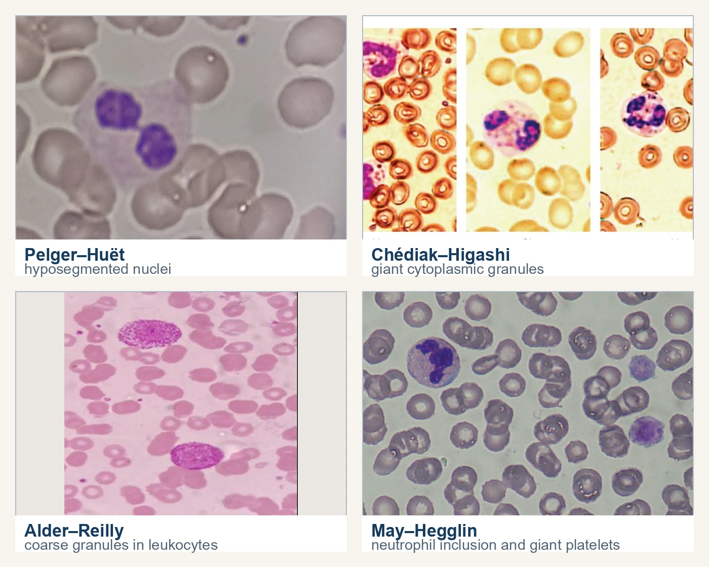
May-Hegglin anomaly and the MYH9 disorder spectrum are autosomal dominant disorders of the nonmuscle myosin heavy chain IIA gene (MYH9) at 22q12.3. The film shows basophilic, Döhle-body-like inclusions in neutrophils, eosinophils, basophils, and monocytes together with giant platelets and variable thrombocytopenia. The inclusions are precipitated myosin heavy chains. True Döhle bodies are remnants of rough endoplasmic reticulum. The MYH9 inclusions are distinguished from them by their presence in several leukocyte lines. Most patients are asymptomatic. In the minority who bleed, bleeding correlates with the degree of thrombocytopenia, and extrahematologic features such as presenile cataracts, nephropathy, and progressive hearing loss can appear over time.5
Inborn errors of immunity with leukocyte findings
In the hematology laboratory, you recognize the disorders below by their count patterns, cell populations, and film clues.1
Scroll sideways to see all columns
| Disorder | Gene | Hematologic and laboratory clues | Recognition notes |
|---|---|---|---|
| Severe combined immunodeficiency, γc (X-linked) type | IL2RG | T and NK lymphocytes nearly absent; B cells present in normal number but nonfunctional | Most common SCID form; symptomatic at 3 to 6 months as maternal immunoglobulin wanes; lymphopenia on the CBC is the clue |
| 22q11.2 deletion syndrome | Recurrent 22q11.2 deletion that includes TBX1 | Variable T-cell deficiency from thymic hypoplasia | Prevalence about 1 in 6,000 births in a US population study, and 1 in 3,800 among Hispanic births; cardiac and palatal findings usually point to the diagnosis first6 |
| X-linked agammaglobulinemia | BTK | Profoundly decreased or absent B cells; all serum immunoglobulin isotypes reduced | Infections often begin after maternal antibody wanes; quantitative immunoglobulins are reduced, and lymphocyte-subset flow cytometry with CD19-based enumeration shows markedly reduced or absent circulating B cells, a result the routine CBC and differential do not provide14 |
| Wiskott-Aldrich syndrome | WAS | T cells decreased; thrombocytopenia with small platelets | Small platelets on the film are the diagnostically important hematologic clue |
| Chronic granulomatous disease | CYBB and other NADPH oxidase genes | Normal counts with defective respiratory burst | About two-thirds of cases are X-linked; dihydrorhodamine flow cytometry assesses the oxidative-burst response, with the assay limitations described below7 |
| Leukocyte adhesion deficiency I | ITGB2 (CD18) | Marked neutrophilia without pus; cells cannot leave the circulation | Severe disease has CD18 expression below 2% of normal and moderate disease 2 to 30%;8 delayed umbilical-cord separation and recurrent infection begin in infancy9 |
| Leukocyte adhesion deficiency II | SLC35C1 | Neutrophilia from defective selectin-ligand fucosylation | Infections are milder than LAD I; growth retardation and coarse facial features accompany it |
| Leukocyte adhesion deficiency III | FERMT3 (kindlin-3) | Mild LAD-I-like infection pattern | Normal integrin expression with failed inside-out activation; platelet GPIIb/IIIa activation also fails, producing Glanzmann-like bleeding |
| Severe congenital and cyclic neutropenia | ELANE can cause either pattern; HAX1 causes an autosomal recessive congenital form | Persistently low counts or recurring nadirs and recovery | Severe congenital neutropenia carries myeloid malignancy risk; cyclic neutropenia is a distinct count pattern without that established association10 |
| Shwachman-Diamond syndrome | SBDS | Marrow failure with cytopenias, myelodysplasia, and leukemia risk | Exocrine pancreatic insufficiency with malabsorption and failure to thrive accompanies the marrow picture11 |
| WHIM syndrome | CXCR4 | Neutropenia, lymphopenia, monocytopenia, hypogammaglobulinemia | Myelokathexis, marrow neutrophil retention with pyknotic degenerative change, despite abundant mature marrow myeloid cells; extensive warts mark the phenotype12 |
Serial absolute neutrophil counts establish whether neutropenia is sustained or cycles through recurring nadirs and recovery. An ELANE variant can occur in either congenital or cyclic neutropenia, so genotype alone cannot demonstrate cycling. A single normal count, low count, or marrow examination captures only one time point. Only a sufficiently long series shows the pattern, and small oscillations can be hard to recognize.10
Dihydrorhodamine (DHR) fluorescence depends on peroxidase activity as well as oxidant generation. Complete myeloperoxidase (MPO) deficiency can markedly reduce the neutrophil DHR response even when oxidase function is preserved. Eosinophils may retain a normal DHR response in this setting. Interpret an abnormal screen against a control handled at the same time, and follow it with appropriate confirmatory studies. When an independent assay shows preserved superoxide production, check MPO as one explanation for the discordance. A low DHR signal alone does not establish chronic granulomatous disease.13
Separating anomaly from reactive and clonal findings
Use the questions below to evaluate an unexpected leukocyte finding. Persistence, family pattern, and the rest of the CBC are the most useful findings:
Scroll sideways to see all columns
| Question | Suggests hereditary anomaly | Suggests reactive or clonal process |
|---|---|---|
| Is the finding stable across serial specimens? | Persists unchanged over months | Varies with illness, treatment, or recovery |
| Do family members share the pattern? | Positive family films support autosomal dominant forms such as Pelger-Huët and May-Hegglin | Negative, or the patient is the only affected member |
| Which cells carry the change? | Multiple leukocyte lines in Pelger-Huët, Alder-Reilly, and May-Hegglin | Toxic granulation stays in neutrophils; pseudo-Pelger-Huët is usually neutrophil-restricted outside MDS |
| Is there infection, inflammation, or a drug explanation? | Absent | Present and quantitatively plausible |
| Are blasts, dysplasia, or other cytopenias present? | Absent | Present in myeloid neoplasia; pseudo-Chédiak-Higashi granules and pseudo-Pelger-Huët both occur there |
Check the platelets as well. Small platelets suggest Wiskott-Aldrich syndrome, giant platelets with leukocyte inclusions suggest the MYH9 spectrum, and dense-granule deficiency contributes to bleeding in Chédiak-Higashi syndrome. The platelet disorder lessons cover those count and function patterns.
Reporting considerations
Report an anomaly as an observed pattern with a comment, and classify the cells correctly. Pelger-Huët cells are counted as segmented neutrophils, and counting them as bands produces a false left shift. When the pattern is unexplained, put suggested next steps in the comment: film review of family members for suspected Pelger-Huët or May-Hegglin anomaly, repeat testing to confirm persistence, and referral to the immune-deficiency pathway for count and function patterns such as sustained lymphopenia in an infant, neutrophilia without pus, or unexplained early-onset neutropenia. Findings in newborns and infants are the most urgent. Delayed cord separation with neutrophilia, giant granules on a film, and small platelets with lymphopenia each call for urgent escalation under your laboratory's notification procedure. Count each cell by its actual type, and use the comment to describe what the pattern might mean.2
Practice
References
- Bousfiha AA, Jeddane L, Moundir A, et al. The 2024 update of IUIS phenotypic classification of human inborn errors of immunity. J Hum Immun. 2025;1(1):e20250002. doi:10.70962/jhi.20250002. Back to text
- Keohane EM, Preston MM, Mirza KM, Walenga JM, eds. Rodak's Hematology: Clinical Principles and Applications. 7th ed. Elsevier; 2025. Accessed August 31, 2026. Back to text
- College of American Pathologists. 2026 Hematology, Clinical Microscopy, and Body Fluids Glossary. 2026:4. Accessed September 7, 2026. Back to text
- Chediak-Higashi syndrome. GeneReviews. Accessed August 31, 2026. Back to text
- MYH9-related disease. GeneReviews. Accessed August 31, 2026. Back to text
- 22q11.2 deletion syndrome. GeneReviews. Accessed August 31, 2026. Back to text
- Chronic granulomatous disease. GeneReviews. Accessed August 31, 2026. Back to text
- Harris ES, Weyrich AS, Zimmerman GA. Lessons from rare maladies: leukocyte adhesion deficiency syndromes. Curr Opin Hematol. 2013;20(1):16-25. doi:10.1097/MOH.0b013e32835a0091. Back to text
- Justiz Vaillant AA, Ahmad F. Leukocyte adhesion deficiency. StatPearls. Updated July 3, 2023. Accessed August 31, 2026. Back to text
- ELANE-related neutropenia. GeneReviews. Accessed August 31, 2026. Back to text
- Shwachman-Diamond syndrome. GeneReviews. Accessed August 31, 2026. Back to text
- Hernandez PA, Gorlin RJ, Lukens JN, et al. Mutations in the chemokine receptor gene CXCR4 are associated with WHIM syndrome, a combined immunodeficiency disease. Nat Genet. 2003;34(1):70-74. doi:10.1038/ng1149. Back to text
- Mauch L, Lun A, O'Gorman MRG, et al. Chronic granulomatous disease (CGD) and complete myeloperoxidase deficiency both yield strongly reduced dihydrorhodamine 123 test signals but can be easily discerned in routine testing for CGD. Clin Chem. 2007;53(5):890-896. doi:10.1373/clinchem.2006.083444. Back to text
- X-linked agammaglobulinemia. GeneReviews. Accessed September 13, 2026. Back to text