When the platelet count is low, first confirm that the count is real, then determine why it is low. The platelet reference interval is approximately 150 to 450 × 103/µL, and thrombocytopenia means a count below the laboratory's lower reference limit. A count below 100 × 103/µL is a separate threshold used in immune thrombocytopenia and several treatment protocols. Clinically significant bleeding is unusual above 50 × 103/µL, risk rises progressively below that level, and a count below 10 × 103/µL carries a high risk of spontaneous severe bleeding.1,2
Purpura describes bleeding into skin or mucosa. Petechiae are small pinpoint lesions. Ecchymoses are larger bruises.15 Three mechanisms produce a true low count: decreased production, increased destruction or consumption, and abnormal distribution or sequestration. Specimen artifact can also produce a falsely low count, and the laboratory can resolve it at the bench.
Confirming the count before classifying it
EDTA-dependent platelet clumping and platelet satellitosis, in which platelets form rosettes around neutrophils, falsely lower the automated count. Clumps counted as white-cell-sized particles can also raise the WBC count, producing pseudoleukocytosis. Room-temperature-reactive platelet autoantibodies contribute to both phenomena. The blood film detects both before an incorrect result is reported, so review the film whenever the count and the film disagree.1
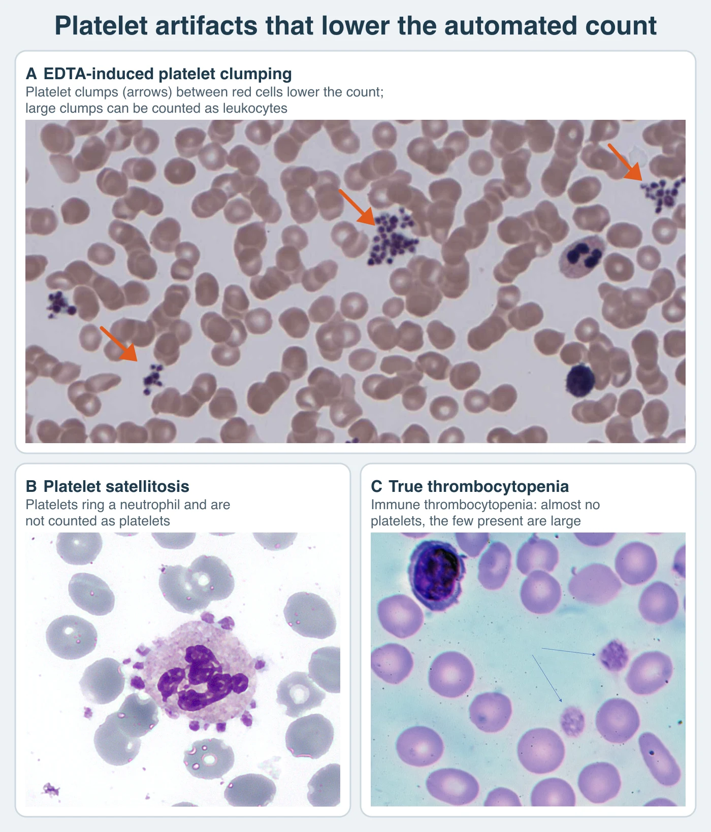
When you suspect EDTA-dependent clumping, request a properly filled citrate tube if your laboratory's validated procedure allows it. Check the new film, because clumping can persist in citrate, and analyze within the validated stability interval. Validate the collection tube and the counting method together. A dilution correction does not correct for persistent clumping.3
Nine parts blood plus one part liquid anticoagulant give a theoretical volume correction of 10/9 = 1.111…. A laboratory procedure may adopt the conventional rounded factor of 1.1 after method verification. For a citrated platelet count of 88 × 103/µL:
Corrected count = citrate count × 10/9 = 88 × 103/µL × 1.111… = 97.8 × 10³/µL
Corrected count = citrate count × 1.1 = 88 × 103/µL × 1.1 = 96.8 × 10³/µL
Both values are shown before the laboratory's reporting round-off. Apply the adopted factor once, and check whether the analyzer or the laboratory information system (LIS) has already applied it. Report other CBC parameters only from a specimen and method suitable for those measurements.3
Decreased production
Congenital causes. Most inherited thrombocytopenias map to specific genes, and estimating platelet size on the film is the fastest way to narrow the differential.1
Scroll sideways to see all columns
| Disorder | Inheritance; gene | Platelet size | Distinguishing feature |
|---|---|---|---|
| Wiskott-Aldrich syndrome | X-linked; WAS | Small | Combined immunodeficiency (see the hereditary leukocyte anomalies lesson) |
| GATA1-related disease | X-linked; GATA1 | Large | Hemolytic anemia; possible congenital dyserythropoietic anemia |
| FLNA-related thrombocytopenia | X-linked; FLNA | Large | Periventricular nodular heterotopia |
| Thrombocytopenia with absent radii | Autosomal recessive; RBM8A | Normal | Bilateral radial aplasia; reduced megakaryocytes; count normalizes into adulthood |
| Bernard-Soulier syndrome, biallelic | Autosomal recessive; GP1BA, GP1BB, or GP9 | Large | Mucocutaneous bleeding (see the qualitative platelet defects lesson)4 |
| Congenital amegakaryocytic thrombocytopenia | Autosomal recessive; MPL | Normal | Marrow aplasia in infancy from defective thrombopoietin-receptor signaling |
| Gray platelet syndrome | Autosomal recessive; NBEAL2 | Giant | Risk of myelofibrosis and splenomegaly |
| Thrombocytopenia with lipid accumulation | Autosomal recessive; ABCG5, ABCG8 | Large | Anemia, tendon xanthomas, atherosclerosis |
| MYH9-related disease | Autosomal dominant; MYH9 | Large | Presenile cataracts, nephropathy, hearing loss; May-Hegglin film picture |
| Paris-Trousseau thrombocytopenia | Autosomal dominant; 11q23.3 deletion | Large | Cardiac and facial defects, developmental delay |
| Bernard-Soulier syndrome, monoallelic | Autosomal dominant; selected GP1BA or GP1BB variants | Large | Often a milder macrothrombocytopenia; bleeding varies4 |
| RUNX1 familial platelet disorder | Autosomal dominant; RUNX1 | Normal | High risk of leukemia or myelodysplastic neoplasm (MDS); qualitative dysfunction often causes easy bruising |
| ANKRD26-related thrombocytopenia | Autosomal dominant; ANKRD26 | Normal | Risk of acute myeloid leukemia (AML) and MDS; identified in about 10% of thrombocytopenic pedigrees in one series5 |
| Congenital radioulnar synostosis with thrombocytopenia | Autosomal dominant; HOXA11 | Normal | Possible evolution to aplastic anemia |
Fanconi anemia, an inherited bone-marrow-failure syndrome, adds thrombocytopenia with bony or visceral abnormalities and progressing pancytopenia.
Acquired causes. Chemotherapeutic agents such as methotrexate, busulfan, cytarabine, cyclophosphamide, and cisplatin commonly cause dose-limiting marrow hypoplasia. Long-term ethanol use can produce persistent severe thrombocytopenia that resolves within weeks of abstinence, sometimes with a rebound thrombocytosis. Interferon, estrogens, chloramphenicol, tranquilizers, and anticonvulsants can suppress megakaryocytopoiesis. Megaloblastic anemia causes ineffective thrombopoiesis: the marrow shows increased, dysplastic megakaryocytes, and the platelets that reach circulation are large with possible reduced survival and function. Viruses including cytomegalovirus (CMV), varicella-zoster virus (VZV), rubella, Epstein-Barr virus (EBV), and dengue suppress production, as do bacterial toxins and direct platelet-bacteria interaction. Meningococcemia is a serious cause of thrombocytopenia. Marrow infiltration by myeloma, lymphoma, metastatic cancer, or fibrosis reduces megakaryocyte numbers directly.1
Increased destruction and consumption
Immune thrombocytopenia
Current terminology defines immune thrombocytopenia (ITP) by duration: newly diagnosed under 3 months, persistent from 3 to 12 months, and chronic beyond 12 months.6
ITP remains a diagnosis of exclusion. Neither the count, platelet size, age, nor a preceding infection confirms it. Review the CBC and film for other cytopenias, abnormal cells, fragmentation, and clumping, and assess possible secondary causes. The autoimmune process can both accelerate platelet destruction and impair production. The course varies at every age. Remission is more common in children, and onset can be abrupt or gradual at any age.2
Immunologic drug-induced thrombocytopenia
Several mechanisms can cause drug-induced immune thrombocytopenia, and only some of them are drug-dependent:
Scroll sideways to see all columns
| Mechanism | Example drugs | Pattern |
|---|---|---|
| Drug-dependent antibody | Quinidine, quinine, sulfonamides; GPIIb/IIIa antagonists | Antibody binds the platelet only when the drug is present; rapid, often severe thrombocytopenia with abrupt bleeding |
| Hapten-induced antibody | Penicillins | Drug plus carrier protein forms a complete antigen; often severe with normal-to-elevated marrow megakaryocytes |
| Drug-induced autoantibody | Gold salts, procainamide, levodopa | Autoantibody persists without continued drug exposure |
| Heparin-induced thrombocytopenia | Heparin | Antibody targets heparin-platelet factor 4 complexes and activates platelets, so thrombocytopenia is accompanied by a high thrombosis risk |
Heparin-induced thrombocytopenia (HIT) is the laboratory-urgent member of this group. In the 4Ts score, the highest-scoring pattern is a platelet count fall greater than 50% with a nadir of at least 20 × 103/µL and a clear onset 5 to 10 days after heparin exposure.7 The main risk is thrombosis. The 4Ts score weighs thrombocytopenia depth, timing, thrombosis, and competing causes to set the pretest probability. Heparin-platelet factor 4 immunoassays are used for screening, and functional assays such as the serotonin-release assay are used for confirmation. When a heparin-exposed patient develops a new thrombocytopenia, flag it promptly, because the risk of thrombosis begins at the same time. The clinical and transfusion services determine anticoagulation and transfusion support.8
Thrombotic microangiopathies
Thrombotic microangiopathy (TMA) combines thrombocytopenia, arteriolar and capillary microthrombi, and microangiopathic hemolytic anemia. The pattern also appears with drugs such as clopidogrel and ticlopidine, transplantation, malignancy, pregnancy, disseminated intravascular coagulation (DIC), catastrophic antiphospholipid syndrome, autoimmune disease, HIV, and malignant hypertension.
Thrombotic thrombocytopenic purpura (TTP). Severe ADAMTS13 deficiency permits ultralarge VWF multimers to sustain platelet-rich microthrombi. TTP may be immune-mediated or congenital. Thrombocytopenia with microangiopathic hemolytic anemia calls for urgent notification even without the classic five clinical findings. Schistocytes, increased lactate dehydrogenase (LDH) and indirect bilirubin, and decreased haptoglobin support the hemolytic pattern. Organ involvement overlaps with other TMAs.9
The prothrombin time (PT) and activated partial thromboplastin time (APTT) are often less disturbed than in overt DIC. Coagulation results cannot reliably distinguish TTP from DIC. ADAMTS13 activity below 10% is the hallmark of TTP. Inhibitor or anti-ADAMTS13 antibody testing helps establish the immune mechanism. Collect the ADAMTS13 specimen before plasma exchange or plasma-containing products when feasible, and communicate the urgent pattern promptly.9
The PLASMIC score estimates the probability of severe ADAMTS13 deficiency while testing is pending. It gives one point each for platelets below 30 × 103/µL; evidence of hemolysis (reticulocytes above 2.5%, undetectable haptoglobin, or indirect bilirubin above 2 mg/dL); no active cancer in the preceding year; no solid-organ or stem-cell transplant; mean cell volume (MCV) below 90 fL; international normalized ratio (INR) below 1.5; and creatinine below 2 mg/dL. A high score supports suspicion of severe ADAMTS13 deficiency. Confirmation depends on the ADAMTS13 result. The score was developed for adults and is less reliable in children and patients with major comorbidities, including pregnancy, cancer, sepsis, or transplantation.9,10
The laboratory communicates the urgent pattern, and the clinical and transfusion services then decide transfusion support in suspected TTP.
Hemolytic uremic syndrome (HUS). Shiga-toxin-associated HUS follows Shigella dysenteriae or enterohemorrhagic E. coli, especially O157:H7, with bloody diarrhea preceding the triad of hemolytic anemia, renal failure, and mild-to-moderate thrombocytopenia. Shiga toxin damages glomerular endothelium, and urinary findings include red cells, protein, and casts. Complement-mediated HUS often involves complement-regulatory protein variants, and secondary forms follow drugs, transplantation, malignancy, pregnancy, and severe hypertension. Prominent kidney injury favors HUS. Neurologic findings and the degree of fragmentation cannot reliably separate it from TTP at presentation. Report the TMA pattern urgently, and state in the report that TTP remains possible until the ADAMTS13 result is available.1
Disseminated intravascular coagulation (DIC). Coagulation activation consumes platelets in intravascular fibrin clots and consumes factors V, VIII, and fibrinogen. Acute DIC produces severe thrombocytopenia, prolonged clotting times, low fibrinogen, and a positive D-dimer. Chronic DIC leaves factors near normal with a moderately low platelet count and increased D-dimer or fibrin degradation products, and it can convert to the acute form. The ISTH overt-DIC score combines the platelet count, D-dimer, PT prolongation, and fibrinogen into a single laboratory decision rule for overt DIC. Interpret the score with its trend and the full clinical context.11
Purpura fulminans. Large, irregular, blue-black hemorrhages progress to tissue necrosis. The laboratory findings resemble DIC, with prolonged clotting times, thrombocytopenia, and hypofibrinogenemia. In neonates it suggests severe congenital protein C or S deficiency. In older children and adults, severe sepsis, especially from Neisseria meningitidis, Streptococcus pneumoniae, or group A or B streptococcus, is the important cause.1
Pregnancy-associated patterns
Gestational thrombocytopenia is the most common cause of thrombocytopenia in pregnancy and is benign once other causes are excluded. Preeclampsia causes thrombocytopenia and can progress to eclampsia, and HELLP syndrome (hemolysis, elevated liver enzymes, low platelets) is a severe variant that is difficult to distinguish from TTP, complement-mediated TMA, HUS, and DIC. ITP remains in the differential at childbearing age, and TTP can recur in pregnancy or postpartum.1
Nonimmune drug and surface effects
Ristocetin, withdrawn from clinical use, agglutinated platelets through VWF-GPIb binding. Hematin, protamine sulfate, and bleomycin can interact with platelets directly and cause transient thrombocytopenia.1
Distribution and dilution
The normal spleen sequesters about one third of the platelet mass, so any splenomegaly, including cirrhosis with portal hypertension, lymphoma, sarcoidosis, and chronic myeloid leukemia (CML), can produce mild thrombocytopenia with a normal total body platelet mass. Hypothermia below about 25 °C during cardiovascular surgery causes transient mild thrombocytopenia and a function defect that reverses on rewarming. Extracorporeal circuits damage and partially activate platelets, occasionally producing severe thrombocytopenia with marked functional impairment. Massive transfusion of stored whole blood dilutes and clears platelets, a rarer problem in the component-therapy era. Mild thrombocytopenia also accompanies chronic renal failure, severe iron deficiency, megaloblastic anemia, and chronic hypoxia.1
Thrombocytosis
Reactive thrombocytosis produces counts of roughly 450 to 800 × 103/µL, occasionally higher, with normal platelet function. Causes include acute blood loss, splenectomy, childbirth, tissue necrosis and surgery, chronic inflammatory disease, infection, exercise, iron deficiency, hemolytic anemia, renal disorders, and malignancy. Production remains responsive to thrombopoietin, the glycoprotein hormone produced mainly by the liver, and the count normalizes as the underlying disorder resolves. Reactive thrombocytosis has a low complication rate.
Sustained, autonomous thrombocytosis is typical of polycythemia vera (PV), CML, primary myelofibrosis (PMF), and essential thrombocythemia (ET). The count alone cannot establish clonality. Exclude reactive causes first. The physical findings, film, marrow, and genetic workup then establish clonality. The myeloid neoplasia lesson gives the full diagnostic criteria for ET from the fifth-edition WHO classification.12
ET arises from clonal proliferation of a multipotent hematopoietic stem cell and requires a sustained count of at least 450 × 103/µL. Three mutually exclusive driver mutations account for most cases: JAK2 V617F in about 60%, CALR in about 25%, and MPL in about 3%. About 8% of patients carry none of the three and are called triple-negative. In a series of 1,000 patients, CALR-mutated ET had higher platelet counts with lower rates of arterial and venous thrombosis at or before diagnosis (7% and 4%) than JAK2-mutated disease (16% and 13%).13 The JAK2 V617F allele burden tends to be higher in PV than in ET, and homozygous clones are more common in PV, but both heterozygous and homozygous patterns occur in either disease. Allele burden alone cannot assign the myeloproliferative neoplasm (MPN) subtype.14
The film can look deceptively benign. Platelets are often normal but can be giant, bizarre, or clumped. Micromegakaryocytes and megakaryocyte fragments appear. Platelets cluster near the thin edge. Anemia is unusual early, and about a third of patients have mild erythrocytosis that must be distinguished from polycythemia vera. Reassess the pattern when marked leukocytosis appears. A platelet count above 400 × 103/µL with a significant marrow erythropoietic or granulopoietic increase suggests PV or PMF. Dyserythropoiesis or dysgranulopoiesis calls for cytogenetic testing for del(5q), inv(3), or t(3;3).
Laboratory findings correlate with the clinical phenotype. Platelets from patients who have thrombosed show increased fibrinogen-binding affinity, elevated thromboxane B2, and elevated β-thromboglobulin, evidence of ongoing in vivo activation. The hemorrhagic phenotype correlates with qualitative defects such as deficient epinephrine receptors and granule abnormalities. Aggregation results in ET are variable and nonspecific: epinephrine nonresponse alone does not separate ET from reactive thrombocytosis.
Urgent laboratory patterns
Scroll sideways to see all columns
| Pattern | Recognize on | Laboratory action |
|---|---|---|
| Suspected TTP or severe TMA | Schistocytes, thrombocytopenia, elevated LDH; PT/APTT may be relatively preserved | Notify urgently; collect the ADAMTS13 specimen before treatment when feasible |
| Suspected HIT | A fall greater than 50% from baseline on heparin, onset 5 to 10 days | Flag promptly; PF4 screening and confirmatory testing per protocol |
| Severe acute DIC | Falling platelets with prolonged clotting times, low fibrinogen, positive D-dimer | Urgent notification; monitor ISTH score trends |
| Spontaneous bleeding risk | Platelet count below 10 × 103/µL, or below 20 with bleeding | Priority handling and critical-value reporting per policy |
Transfusion support in these situations follows blood banking guidance and the institution's transfusion service. The hemostasis lessons describe the procedures behind coagulation testing. In each of these patterns, first confirm the count on the film, then decide how urgently to communicate the result.1
Practice
References
- Keohane EM, Preston MM, Mirza KM, Walenga JM, eds. Rodak's Hematology: Clinical Principles and Applications. 7th ed. Elsevier; 2025. Accessed August 31, 2026. Back to text
- Neunert C, Terrell DR, Arnold DM, et al. American Society of Hematology 2019 guidelines for immune thrombocytopenia. Blood Adv. 2019;3(23):3829-3866. doi:10.1182/bloodadvances.2019000966. Back to text
- Baccini V, Geneviève F, Jacqmin H, et al. Platelet counting: ugly traps and good advice. Proposals from the French-Speaking Cellular Hematology Group (GFHC). J Clin Med. 2020;9(3):808. doi:10.3390/jcm9030808. Back to text
- National Library of Medicine. Bernard-Soulier syndrome. MedlinePlus Genetics. Accessed September 7, 2026. Back to text
- ANKRD26-related thrombocytopenia. GeneReviews. Accessed August 31, 2026. Back to text
- Rodeghiero F, Stasi R, Gernsheimer T, et al. Standardization of terminology, definitions and outcome criteria in immune thrombocytopenic purpura of adults and children: report from an international working group. Blood. 2009;113(11):2386-2393. doi:10.1182/blood-2008-07-162503. Back to text
- Cuker A, Gimotty PA, Crowther MA, Warkentin TE. Predictive value of the 4Ts scoring system for heparin-induced thrombocytopenia: a systematic review and meta-analysis. Blood. 2012;120(20):4160-4167. doi:10.1182/blood-2012-07-443051. Back to text
- Cuker A, Arepally GM, Chong BH, et al. American Society of Hematology 2018 guidelines for management of venous thromboembolism: heparin-induced thrombocytopenia. Blood Adv. 2018;2(22):3360-3392. doi:10.1182/bloodadvances.2018024489. Back to text
- Zheng XL, Vesely SK, Cataland SR, et al. ISTH guidelines for the diagnosis of thrombotic thrombocytopenic purpura. J Thromb Haemost. 2020;18(10):2486-2495. doi:10.1111/jth.15006. Back to text
- Bendapudi PK, Hurwitz S, Fry A, et al. Derivation and external validation of the PLASMIC score for rapid assessment of adults with thrombotic microangiopathies: a cohort study. Lancet Haematol. 2017;4(4):e157-e164. doi:10.1016/S2352-3026(17)30026-1. Back to text
- Toh CH, Hoots WK. The scoring system of the Scientific and Standardisation Committee on Disseminated Intravascular Coagulation of the International Society on Thrombosis and Haemostasis: a 5-year overview. J Thromb Haemost. 2007;5(3):604-606. doi:10.1111/j.1538-7836.2007.02313.x. Back to text
- Tefferi A, Gangat N, Loscocco GG, et al. Essential thrombocythemia: 2024 update on diagnosis, risk stratification, and management. Am J Hematol. 2024;99(4):697-718. doi:10.1002/ajh.27216. Back to text
- Gangat N, Karrar O, Al-Kali A, et al. One thousand patients with essential thrombocythemia: the Mayo Clinic experience. Blood Cancer J. 2024;14(1):11. doi:10.1038/s41408-023-00972-x. Back to text
- Vannucchi AM, Antonioli E, Guglielmelli P, et al. Clinical profile of homozygous JAK2 617V>F mutation in patients with polycythemia vera or essential thrombocythemia. Blood. 2007;110(3):840-846. doi:10.1182/blood-2006-12-064287. Back to text
- DermNet. Purpura. Accessed September 8, 2026. Back to text