On the bench, a myeloid neoplasm often first appears as circulating blasts, a broad left shift with basophilia, or a count that stays high without a reactive cause. Myeloid neoplasms arise from clonal hematopoietic cells and can involve one or several myeloid lineages. The CBC and blood film establish the circulating pattern. Bone marrow morphology, immunophenotyping, cytogenetics, and molecular results then define the disease and explain findings that morphology cannot resolve alone.1,2
Leukemia primarily involves marrow and blood, although tissue masses can occur. Lymphoma primarily forms tissue masses and may enter a leukemic phase. Acute disease usually shows blasts and rapid loss of normal hematopoiesis. Chronic disease usually retains more maturation and can present with markedly increased mature cells. Exceptions occur within these patterns: acute leukemia can have a low or normal WBC count, and a chronic neoplasm can contain immature cells.1,3
Classification here follows the fifth edition of the World Health Organization classification (WHO5). The International Consensus Classification (ICC) differs at selected blast thresholds and in several disease names, so state in each report which classification you applied.1,4,5
Integrated laboratory classification
The diagnostic record includes prior counts, cytotoxic therapy, occupational or environmental exposures, family history, known germline predisposition, and an earlier myelodysplastic neoplasm (MDS), myelodysplastic/myeloproliferative neoplasm (MDS/MPN), or myeloproliferative neoplasm (MPN). These facts can become formal disease qualifiers once the morphologic and genetic criteria for the named neoplasm are met.1
Scroll sideways to see all columns
| Evidence | Main contribution | Limitation |
|---|---|---|
| CBC and blood film | Detects cytopenias, blasts, dysplasia, left shift, basophilia, monocytosis, eosinophilia, erythrocytosis, thrombocytosis, and leukoerythroblastosis | A normal or low WBC count does not exclude acute leukemia. A reactive pattern can resemble a clonal process. |
| Marrow aspirate | Supports blast enumeration, lineage morphology, dysplasia assessment, iron and ring-sideroblast review, and specimen allocation | Hemodilution or a dry tap can make the aspirate unrepresentative. |
| Marrow core biopsy | Shows cellularity, architecture, fibrosis, clustered or atypical megakaryocytes, mast-cell aggregates, and focal disease | Decalcification and sampling can limit some studies. Reserve fresh material when possible. |
| Flow cytometry | Assigns lineage, identifies an abnormal population, and supports acute leukemia classification | Antigen patterns require the full panel and morphologic context. A single marker rarely defines a disease. |
| Karyotype and fluorescence in situ hybridization (FISH) | Detects balanced rearrangements, copy-number changes, clonal evolution, and selected cryptic targets | A normal karyotype does not exclude a cryptic fusion or sequence variant. |
| Reverse-transcription polymerase chain reaction (RT-PCR) and sequencing | Confirms fusions or mutations that define a category and establishes targets for later measurement when applicable | Assay coverage and transcript design determine what can be detected. A negative limited panel leaves untested abnormalities unresolved. |
When acute leukemia is suspected, allocate fresh blood and marrow early, because morphology, flow cytometry, chromosome analysis, FISH, rapid fusion testing, and broader sequencing may all use the same collection.2,3
Acute myeloid leukemia
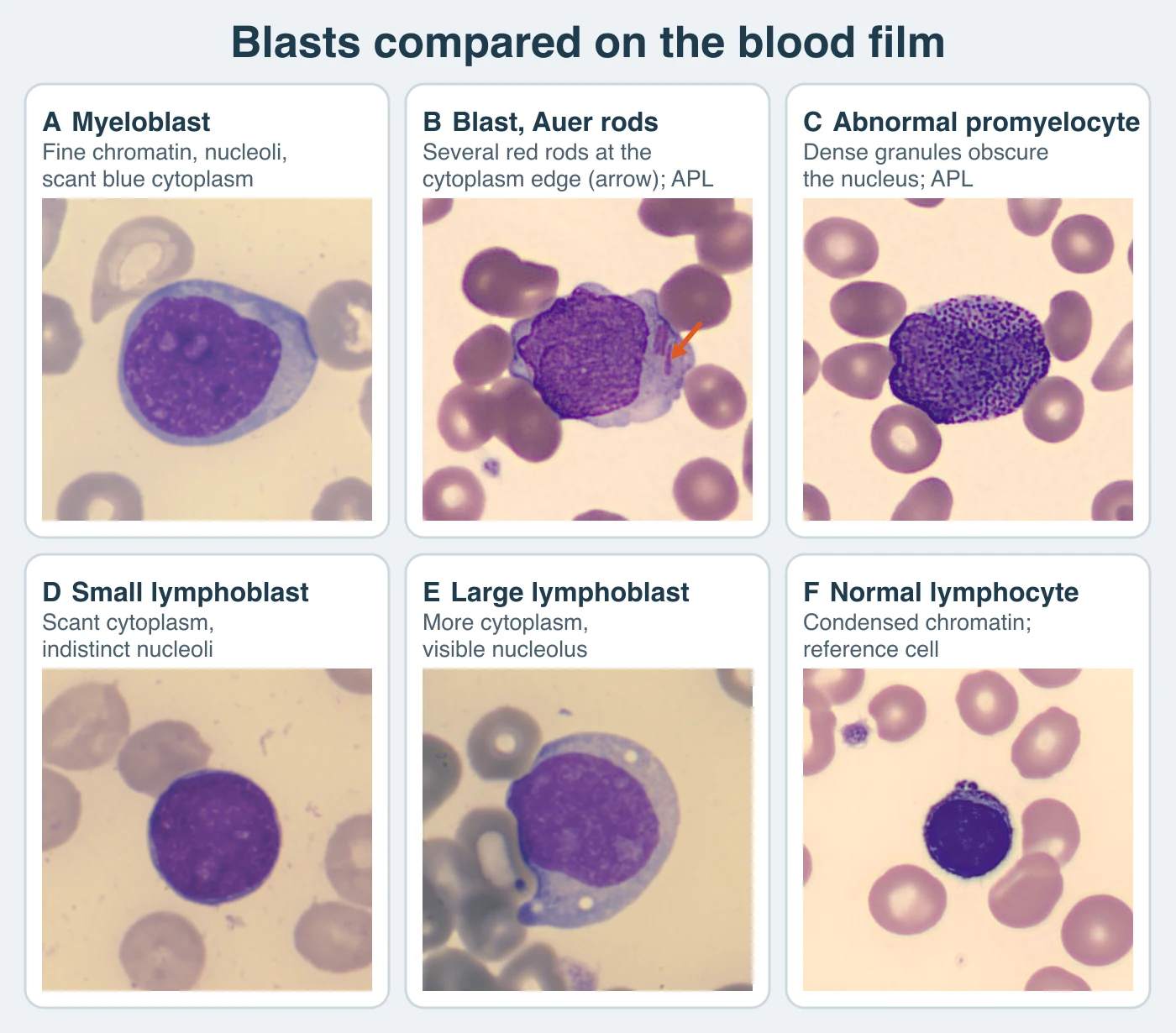
Acute myeloid leukemia (AML) replaces normal marrow with a clonal blast population. Anemia, thrombocytopenia, and neutropenia can occur together even when the total WBC count is high. The blood film may show circulating blasts, Auer rods, or dysplastic maturing cells. A myeloblast has fine chromatin, one or more nucleoli, and scant blue cytoplasm. An Auer rod is a red, needle-shaped cytoplasmic inclusion. The abnormal promyelocyte of acute promyelocytic leukemia (APL) is packed with dense granules that can obscure the nucleus. Marrow examination establishes blast burden and morphology. Flow cytometry confirms lineage and identifies the abnormal phenotype. Cytogenetic and molecular findings now define many AML categories.1,2,3
Suspected acute promyelocytic leukemia is a medical emergency. Abnormal promyelocytes, multiple Auer rods, thrombocytopenia, prolonged clotting times, low fibrinogen, or increased D-dimer call for immediate notification under your laboratory's urgent-result procedure, because the coagulopathy can be severe. Rapid PML::RARA testing and coagulation studies proceed without waiting for the complete leukemia workup.2,6
High tumor burden can produce spontaneous tumor lysis or increase the risk after cytotoxic therapy. Using the Howard refinement, laboratory tumor lysis syndrome requires at least two qualifying abnormalities in the same 24-hour period, from 3 days before through 7 days after treatment. Adult thresholds are uric acid above 8 mg/dL, phosphorus above 4.5 mg/dL, potassium above 6 mmol/L, or corrected calcium below 7 mg/dL. The pediatric uric acid threshold follows the age-specific upper limit, and phosphorus must exceed 6.5 mg/dL. This refinement excludes a 25% change from baseline as a stand-alone criterion. Do not mix it with the original Cairo–Bishop change criterion. Clinical tumor lysis adds qualifying kidney injury or clinical complications such as arrhythmia or seizure. Escalation depends on how fast the results are changing and on the local critical-value policy.7
WHO5 genetic categories
WHO5 classifies AML first by defining genetic abnormalities. Most genetically defined AML types have no fixed minimum blast percentage when the lesion is present and the clinicopathologic findings support AML. AML with BCR::ABL1 and AML with a qualifying CEBPA mutation still require at least 20% blasts. AML, myelodysplasia-related (AML-MR), and AML defined by differentiation retain their stated 20% requirements. AML with other defined genetic alterations requires increased blasts and clinicopathologic support without a fixed WHO5 minimum.1
Scroll sideways to see all columns
| WHO5 category | Defining evidence | Blast rule and laboratory implication |
|---|---|---|
| AML with PML::RARA | Defining fusion | No fixed minimum when the clinicopathologic findings support the diagnosis. Rapid testing is urgent because coagulopathy can be severe. |
| AML with RUNX1::RUNX1T1 or CBFB::MYH11 | Core-binding-factor fusion | No fixed minimum. Morphology can suggest the category. The fusion result establishes it. |
| AML with DEK::NUP214 or RBM15::MRTFA | Defining fusion | No fixed minimum. Cytogenetic or molecular confirmation is required. |
| AML with KMT2A, MECOM, or NUP98 rearrangement | Defining rearrangement | A count below 20% is accepted. Some rearrangements are cryptic on conventional karyotyping. |
| AML with NPM1 mutation | Defining mutation in the appropriate setting | No fixed minimum. Clinicopathologic correlation remains required. |
| AML with BCR::ABL1 | Defining fusion | At least 20% blasts. The threshold helps separate AML from CML, and the full record may still be needed to resolve that distinction. |
| AML with CEBPA mutation | Biallelic mutation or a qualifying basic leucine zipper-region mutation | At least 20% blasts. Describe the detected variant pattern in the laboratory report. |
| AML-MR | At least 20% blasts plus a specified MDS-related cytogenetic or molecular abnormality and/or a qualifying history of MDS or MDS/MPN | Morphologic dysplasia alone does not establish AML-MR. The eight WHO5 MDS-related genes are SRSF2, SF3B1, U2AF1, ZRSR2, ASXL1, EZH2, BCOR, and STAG2. |
| AML with other defined genetic alterations | A recognized genetic alteration that falls outside the named rows | No fixed minimum. Increased blasts and clinicopathologic correlation are required, and the report states the abnormality and classification authority. |
ICC generally requires at least 10% blasts for genetically defined AML, including AML with PML::RARA, NPM1 mutation, and an in-frame basic leucine zipper-region CEBPA mutation. ICC retains 20% for AML with BCR::ABL1 and uses an MDS/AML category for many myelodysplasia-related or TP53-mutated cases with 10% to 19% blasts. If you apply these ICC thresholds to a WHO5 diagnosis, name the framework change in the report.5
Post-cytotoxic therapy and an associated germline pathogenic variant are qualifiers attached to the most specific disease name. AML arising from an MPN remains classified with the MPN. A previous MDS or MDS/MPN can satisfy the history component of AML-MR when the other AML requirements are met.1
Differentiation patterns
When a defining genetic category and AML-MR do not apply, WHO5 classifies AML by differentiation. These types generally require at least 20% blasts. French-American-British (FAB) M0 through M7 terms remain useful historical shorthand, especially for morphology and cytochemistry. The table below uses the current names, which describe the differentiation pattern.1,3
Scroll sideways to see all columns
| Differentiation pattern | Morphologic and immunophenotypic basis |
|---|---|
| Minimal differentiation | Blasts lack convincing morphologic or cytochemical myeloid differentiation. An appropriate immunophenotypic panel establishes myeloid lineage. |
| Without maturation | Myeloid differentiation is present and maturing granulocytic cells account for less than 10% of nonerythroid cells. |
| With maturation | Maturing granulocytic cells account for at least 10%, and monocytic cells and precursors remain below 20%. Auer rods can support the pattern. |
| Acute basophilic leukemia | Blasts and immature or mature basophils show metachromasia with toluidine blue. Blasts lack cytochemical myeloperoxidase, Sudan Black B, and nonspecific esterase, and strong CD117 expression should be absent to exclude mast cell leukemia. |
| Myelomonocytic | Granulocytic and monocytic components each account for at least 20%. The abnormal eosinophil pattern formerly called M4Eo is commonly associated with CBFB::MYH11. |
| Monocytic | Monocytes and their precursors account for at least 80% of nonerythroid cells. Monoblasts and promonocytes are included in blast assessment. |
| Acute erythroid leukemia | Erythroid cells usually occupy at least 80% of marrow elements and proerythroblasts at least 30%. Biallelic TP53 alteration is common, and this diagnosis supersedes AML-MR. |
| Acute megakaryoblastic leukemia | At least 20% blasts show convincing megakaryocytic differentiation, commonly supported by CD41, CD61, CD42b, or von Willebrand factor. Defined genetic and Down-syndrome-associated entities are excluded first. |
Related precursor and tissue presentations
Scroll sideways to see all columns
| Entity | Laboratory recognition |
|---|---|
| Myeloid sarcoma | A tissue mass composed of myeloid blasts requires biopsy morphology and immunophenotyping. Blood and marrow studies determine whether AML, MDS, MDS/MPN, or MPN is present elsewhere. |
| Transient abnormal myelopoiesis | In an infant with constitutional trisomy 21, a clonal blast proliferation with a somatic exon 2 or 3 GATA1 mutation can mimic AML. WHO5 confines this diagnosis to the first 6 months of life, and most cases regress spontaneously during early infancy. Myeloid leukemia associated with Down syndrome occurs later in childhood and commonly has megakaryoblastic differentiation. |
| Blastic plasmacytoid dendritic-cell neoplasm | Cells commonly express CD123 with a plasmacytoid dendritic-cell marker such as CD303, CD304, TCF4, or TCL1, often with CD4 or CD56. They lack lineage-defining MPO and surface or cytoplasmic CD3. Skin, marrow, blood, lymph nodes, and the central nervous system can be involved. |
| Acute leukemia of ambiguous lineage | Lineage-defining morphology and flow results leave one lineage unresolved or support more than one. WHO5 genetic groups include mixed-phenotype acute leukemia with BCR::ABL1, KMT2A rearrangement, or ZNF384 rearrangement and acute leukemia of ambiguous lineage with BCL11B rearrangement. Immunophenotypically defined groups include mixed-phenotype B/myeloid, T/myeloid, and rare types, acute leukemia of ambiguous lineage not otherwise specified, and acute undifferentiated leukemia. |
Interpret a tissue diagnosis, a limited antigen panel, or a single fusion result with the blood, marrow, and complete lineage assessment.1,8,9
Myelodysplastic neoplasms
On the bench, MDS often appears as unexplained cytopenias with macrocytosis, hypogranular or hypolobated neutrophils, or giant platelets on the film. Myelodysplastic neoplasms (MDS) are clonal myeloid neoplasms defined by cytopenia with morphologic dysplasia and/or a defining genetic abnormality. Marrow is often normocellular or hypercellular, but hypoplastic MDS is a recognized type. Dysplasia must involve at least 10% of a lineage when it is used as a diagnostic feature. No single dysplastic change is specific.1,10
Scroll sideways to see all columns
| Lineage | Common dysplastic findings |
|---|---|
| Erythroid | Nuclear budding, internuclear bridges, multinuclearity, irregular nuclear contours, megaloblastoid change, abnormal hemoglobinization, and ring sideroblasts |
| Granulocytic | Hypogranulation, abnormal nuclear segmentation, pseudo-Pelger-Huët forms, abnormal chromatin clumping, and increased blasts |
| Megakaryocytic | Micromegakaryocytes, separated nuclear lobes, hypolobation, and abnormal clustering |
| Peripheral blood | Macrocytosis, cytopenias, hypogranular or hypolobated neutrophils, giant or hypogranular platelets, nucleated red cells, and circulating blasts |
Vitamin B12 or folate deficiency, copper deficiency, zinc excess, heavy-metal exposure, medications, cytotoxic therapy, HIV and other infections, parvovirus, paroxysmal nocturnal hemoglobinuria, aplastic anemia, Fanconi anemia, and other germline marrow-failure disorders can produce cytopenia or dysplasia. The diagnosis requires exclusion of a convincing mimic and correlation with cytogenetic and molecular evidence. A trephine core is especially important in a hypocellular or fibrotic specimen.3,10
WHO5 MDS groups
Adult WHO5 groups use the blast ranges below together with group-specific morphologic, genetic, and exclusion requirements.1,10
Scroll sideways to see all columns
| WHO5 group | Blood blasts | Marrow blasts | Defining feature |
|---|---|---|---|
| MDS with low blasts and isolated 5q deletion | Less than 2% | Less than 5% | del(5q) alone or with one additional abnormality other than monosomy 7 or del(7q); compatible megakaryocytic morphology |
| MDS with low blasts and SF3B1 mutation | Less than 2% | Less than 5% | SF3B1 mutation with the permitted cytogenetic context; at least 15% ring sideroblasts can substitute for the mutation in the accepted related category |
| MDS with biallelic TP53 inactivation | Less than 20% | Less than 20% | Two TP53 mutations or one mutation with deletion or copy-neutral loss of heterozygosity; this category supersedes MDS-5q and MDS-SF3B1 |
| MDS with low blasts | Less than 2% | Less than 5% | Cytopenia and qualifying dysplasia after exclusions |
| MDS, hypoplastic | Less than 2% | Less than 5% | Age-adjusted marrow cellularity of 25% or less with MDS evidence and exclusion of aplastic anemia, PNH, drugs, toxins, nutritional causes, and germline marrow failure |
| MDS with increased blasts 1 | 2% to 4% | 5% to 9% | Increased blasts within either stated range |
| MDS with increased blasts 2 | 5% to 19% | 10% to 19% | Either range or the presence of Auer rods |
| MDS with fibrosis | 2% to 19% | 5% to 19% | Significant marrow fibrosis, generally grade 2 to 3, with MDS morphology |
WHO5 retains 20% blasts as the usual MDS-to-AML boundary. Most AML types with defining genetic abnormalities can cross that boundary below 20%. ICC instead uses an MDS/AML category for many cases with 10% to 19% blasts. State the framework in the report, and record the measured blood and marrow blast percentages.1,5
MDS/MPN overlap disorders
Scroll sideways to see all columns
| Entity | Core laboratory pattern | Required context and exclusions |
|---|---|---|
| Chronic myelomonocytic leukemia (CMML) | Persistent absolute monocytes at least 0.5 × 10³/µL and at least 10% of leukocytes; fewer than 20% blasts, including monoblasts and promonocytes | At least one supporting criterion is required when monocytes are at least 1.0 × 10³/µL. At 0.5 to less than 1.0 × 10³/µL, dysplasia and a clonal cytogenetic or molecular abnormality are required. Classical monocytes above 94% can support the diagnosis. CML, other MPNs, myeloid/lymphoid neoplasm (MLN) with tyrosine-kinase fusions, and reactive causes are excluded. |
| Myelodysplastic CMML | CMML criteria with WBC below 13 × 103/µL | The WBC split describes the dysplastic versus proliferative presentation. |
| Myeloproliferative CMML | CMML criteria with WBC at least 13 × 103/µL | Persistent neutrophilia can accompany the monocytosis. |
| MDS/MPN with neutrophilia | WBC at least 13 × 103/µL, at least 10% immature granulocytes, clear dysgranulopoiesis, and fewer than 20% blasts | BCR::ABL1, MLN with tyrosine-kinase fusions, other MPNs, and other MDS/MPNs are excluded. SETBP1 or ETNK1 supports the diagnosis. JAK2, CALR, MPL, or CSF3R calls for reassessment for another process. The historical name is atypical CML. |
| MDS/MPN with SF3B1 mutation and thrombocytosis | Anemia, platelets at least 450 × 103/µL, blood blasts below 1%, marrow blasts below 5%, SF3B1 mutation, and erythroid dysplasia | The accepted related term MDS/MPN with ring sideroblasts and thrombocytosis applies to qualifying SF3B1-wild-type cases with at least 15% ring sideroblasts. |
| MDS/MPN, not otherwise specified | Mixed dysplastic and proliferative findings that meet the category criteria | This is a diagnosis of exclusion after defined MDS, MPN, MDS/MPN, and reactive disorders have been addressed. |
WHO5 uses persistent monocytosis without assigning a fixed minimum interval in its core CMML table. Repeat CBCs document that the monocytosis is sustained while transient reactive causes are investigated.1,10
Myeloproliferative neoplasms
Myeloproliferative neoplasms (MPNs) produce one or more mature myeloid lineages and often preserve a full maturation sequence. A high count can mask ineffective hematopoiesis, evolving fibrosis, or an increasing blast population. Blood-film comparison, marrow architecture, and the defining genetic finding distinguish the major groups.1,3
Chronic myeloid leukemia
CML commonly produces marked neutrophilic leukocytosis with myelocytes, metamyelocytes, and other granulocytic stages, accompanied by basophilia and sometimes eosinophilia. Platelets may be increased. The marrow is hypercellular with granulocytic proliferation and relatively small hypolobated megakaryocytes. Nucleated red cells are usually sparse until the disease is advanced or fibrotic. The benign leukocyte disorders lesson compares this pattern with a leukemoid reaction and explains the historical LAP score.
CML is established by detecting BCR::ABL1. Conventional cytogenetics, FISH, or molecular testing can confirm the fusion. A visible t(9;22) is not required because cryptic and variant rearrangements occur. Marrow morphology and karyotyping help assess phase and additional chromosomal abnormalities. Baseline RT-PCR identifies the transcript for later quantitative measurement. A limited assay for common p210 or p190 transcripts can miss an atypical fusion.11,12
WHO5 uses chronic phase and blast phase without a separate formal accelerated phase, and treats 10% to 19% blasts, basophils at 20% or more, additional clonal abnormalities, or treatment resistance as high-risk chronic-phase findings. The 2025 European LeukemiaNet recommendations compare the WHO5 and ICC phase definitions without fully endorsing either and list the blast and basophil findings as high-risk features at diagnosis. Blast phase is defined by at least 20% myeloid blasts in blood or marrow, an extramedullary blast proliferation, or increased lymphoblasts in blood or marrow. The lymphoblast cutoff is not established, so report bona fide lymphoblasts urgently. ICC retains an accelerated-phase category, so state which framework the phase terminology follows.1,5,12
Polycythemia vera and classic MPN patterns
WHO5 polycythemia vera (PV) criteria use three major findings: hemoglobin above 16.5 g/dL in men or 16.0 g/dL in women, or hematocrit above 49% in men or 48% in women; marrow panmyelosis with trilineage proliferation; and a JAK2 V617F or exon 12 mutation. A subnormal serum erythropoietin concentration is the minor criterion. Diagnosis requires all three major criteria or the first two major criteria plus the minor criterion.1 The erythrocytosis lesson covers secondary hypoxic, drug-associated, erythropoietin-producing, and relative causes.1,13
Scroll sideways to see all columns
| Neoplasm | Blood pattern | Marrow pattern | Defining or supporting genetics |
|---|---|---|---|
| CML | Neutrophilic leukocytosis with a broad left shift and basophilia | Granulocytic hypercellularity with small hypolobated megakaryocytes | BCR::ABL1 |
| PV | Erythrocytosis, often with leukocytosis or thrombocytosis | Panmyelosis with pleomorphic mature megakaryocytes | JAK2 V617F or exon 12 mutation |
| Essential thrombocythemia | Persistent platelets at least 450 × 103/µL | Proliferation of large, mature megakaryocytes with hyperlobulated nuclei and little granulocytic or erythroid left shift | A JAK2, CALR, or MPL mutation is the genetic major criterion. In a driver-negative case, another clonal marker or absence of evidence for reactive thrombocytosis is a minor criterion. |
| Prefibrotic primary myelofibrosis | Anemia or leukocytosis can occur; leukoerythroblastosis is less typical than in overt disease | Megakaryocytic proliferation and atypia, fibrosis grade 0 to 1, increased age-adjusted cellularity with granulocytic proliferation, and often decreased erythropoiesis | JAK2, CALR, MPL, or another clonal marker |
| Overt primary myelofibrosis | Anemia, leukoerythroblastosis, dacryocytes, abnormal platelets, and circulating immature myeloid cells | Atypical clustered megakaryocytes with fibrosis grade 2 to 3; aspiration can produce a dry tap | JAK2, CALR, MPL, or another clonal marker |
Essential thrombocythemia (ET) requires all four major criteria: platelets at least 450 × 103/µL, the characteristic marrow pattern, exclusion of other defined myeloid neoplasms, and a JAK2, CALR, or MPL mutation. When the driver mutation is absent, the first three major criteria plus the minor clonality or reactive-cause criterion can establish the diagnosis. The quantitative platelet disorders lesson covers its full distinction from reactive thrombocytosis, prefibrotic primary myelofibrosis, PV, and CML.1,14
Primary myelofibrosis (PMF) has separate prefibrotic and overt fibrotic forms. Each requires all three major criteria: the form-specific megakaryocytic and marrow pattern; exclusion of CML, PV, ET, MDS, and another myeloid neoplasm; and JAK2, CALR, MPL, another clonal marker, or convincing evidence against reactive fibrosis. At least one form-appropriate minor criterion is also required. Minor findings include unexplained anemia, WBC at least 11 × 103/µL, palpable splenomegaly, serum lactate dehydrogenase above the local upper limit, and, for overt PMF, leukoerythroblastosis. Confirm applicable minor findings on serial assessment.1,15
Other proliferative and related myeloid neoplasms
Several eosinophilic, tyrosine-kinase-fusion, mast-cell, and pediatric myeloid neoplasms fall outside the classic CML, PV, ET, and PMF group in WHO5. Overlapping CBC patterns make exclusion testing necessary for classification.1
Scroll sideways to see all columns
| Entity | Core criteria and interpretation |
|---|---|
| Chronic neutrophilic leukemia | WBC at least 25 × 103/µL; segmented neutrophils plus bands at least 80%; immature granulocytes below 10%; blood blasts usually below 2%; monocytes below 10% and below 1.0 × 10³/µL; no significant dysgranulopoiesis; hypercellular neutrophilic marrow with blasts below 5%; and exclusion of a reactive cause or another myeloid neoplasm. An activating CSF3R mutation supports most cases. A mutation-negative case requires persistent neutrophilia, clonality, splenomegaly, and a rigorous exclusion of reactive causes. |
| Chronic eosinophilic leukemia (CEL) | Eosinophils above 1.5 × 10³/µL on at least two examinations over at least 4 weeks, with both evidence of clonality and abnormal marrow morphology. Reactive eosinophilia, another MPN or MDS/MPN, mastocytosis, AML, and a myeloid/lymphoid neoplasm with a tyrosine-kinase fusion are excluded. WHO5 uses CEL. ICC uses CEL, NOS. |
| Myeloid/lymphoid neoplasm with eosinophilia and tyrosine-kinase gene fusion | Defined by a rearrangement involving PDGFRA, PDGFRB, FGFR1, PCM1::JAK2, or another recognized tyrosine kinase such as ETV6::ABL1 or selected FLT3 rearrangements. Eosinophilia is common, although its absence does not exclude this category. The cryptic FIP1L1::PDGFRA fusion can have a normal karyotype and require targeted FISH, RT-PCR, or sequencing. |
| Idiopathic hypereosinophilic syndrome | Hypereosinophilic syndrome in the broad sense is persistent hypereosinophilia with eosinophil-attributable organ damage, classified further by cause as reactive, neoplastic, familial, or idiopathic. The idiopathic form is diagnosed after reactive, familial, lymphocyte-variant, CEL, tyrosine-kinase-fusion, and other defined neoplastic causes have been excluded. Hypereosinophilia without attributable organ injury does not meet the syndrome definition. |
| Juvenile myelomonocytic leukemia | An early-childhood RAS-pathway MPN with persistent monocytosis and disease-specific clinical, morphologic, and molecular criteria. It is classified with MPNs in WHO5. |
| MPN, not otherwise specified | A persistent myeloproliferative pattern that fails to meet criteria for a defined MPN after reactive causes and other myeloid neoplasms have been excluded. State in the report the findings that support clonality and the criteria that remain unmet. |
The chronic neutrophilic leukemia (CNL) row uses the WHO5 WBC threshold. ICC permits a threshold of at least 13 × 103/µL when an activating CSF3R mutation is present and retains at least 25 × 103/µL when it is absent.1,5
WHO5 recognizes cutaneous mastocytosis, systemic mastocytosis, and mast cell sarcoma. Cutaneous mastocytosis is confined to skin under the category criteria. Mast cell sarcoma is a destructive tissue tumor that requires histologic and immunophenotypic confirmation.1,16
Systemic mastocytosis
The major criterion for systemic mastocytosis is multifocal dense aggregates of at least 15 mast cells in marrow or another extracutaneous organ. The four minor criteria are:
- at least 25% spindle-shaped or otherwise atypical mast cells in the relevant tissue;
- an activating KIT mutation;
- aberrant CD2, CD25, or CD30 expression on mast cells; and
- baseline serum tryptase above 20 ng/mL, adjusted for hereditary alpha-tryptasemia and omitted when an associated myeloid neoplasm explains the increase.
Diagnosis requires the major criterion plus at least one minor criterion, or at least three minor criteria. WHO5 systemic types are bone marrow, indolent, smoldering, aggressive, systemic mastocytosis with an associated hematologic neoplasm, and mast cell leukemia. Mast cell leukemia requires mast cells to occupy at least 20% of the marrow aspirate or smear in the appropriate cytologic setting.1,16
Integrated reporting
An integrated report records blood and marrow blast percentages, specimen limitations, relevant dysplasia and fibrosis, lineage assignment, the defining fusion or mutation, the classification framework, and any qualifying history. Handle suspected APL, a rapidly increasing blast population, or critical coagulation and tumor-lysis results under your laboratory's urgent-notification procedure.
State discordant results in the interpretation, such as a dry tap with a diagnostic core, a normal karyotype with a cryptic fusion, apparent reactive neutrophilia with a clonal CSF3R result, or MDS-range blasts with an AML-defining lesion. Explain how the evidence was reconciled and which assay limitations remain. Record each finding, and describe in plain words how the findings fit together.1,2
Practice
References
- Khoury JD, Solary E, Abla O, et al. The 5th edition of the World Health Organization Classification of Haematolymphoid Tumours: myeloid and histiocytic/dendritic neoplasms. Leukemia. 2022;36(7):1703-1719. doi:10.1038/s41375-022-01613-1. Back to text
- Arber DA, Borowitz MJ, Cessna M, et al. Initial diagnostic workup of acute leukemia: guideline from the College of American Pathologists and the American Society of Hematology. Arch Pathol Lab Med. 2017;141(10):1342-1393. doi:10.5858/arpa.2016-0504-CP. Back to text
- Keohane EM, Preston MM, Mirza KM, Walenga JM, eds. Rodak's Hematology: Clinical Principles and Applications. 7th ed. Elsevier; 2025. Accessed August 30, 2026. Back to text
- International Agency for Research on Cancer. WHO Classification of Tumours: haematolymphoid tumours, current classification structure. Accessed August 30, 2026. Back to text
- Arber DA, Orazi A, Hasserjian RP, et al. International Consensus Classification of myeloid neoplasms and acute leukemias: integrating morphologic, clinical, and genomic data. Blood. 2022;140(11):1200-1228. doi:10.1182/blood.2022015850. Back to text
- Sanz MA, Fenaux P, Tallman MS, et al. Management of acute promyelocytic leukemia: updated recommendations from an expert panel of the European LeukemiaNet. Blood. 2019;133(15):1630-1643. doi:10.1182/blood-2019-01-894980. Back to text
- Howard SC, Jones DP, Pui CH. The tumor lysis syndrome. N Engl J Med. 2011;364(19):1844-1854. doi:10.1056/NEJMra0904569. Back to text
- National Cancer Institute. Myeloid proliferations related to Down syndrome treatment (PDQ), health professional version. Accessed August 30, 2026. Back to text
- Lee YJ, Kim Y, Park SH, Jo JC. Plasmacytoid dendritic cell neoplasms. Blood Res. 2023;58(Suppl 1):S90-S95. doi:10.5045/br.2023.2023052. Back to text
- Hasserjian RP, Germing U, Malcovati L. Diagnosis and classification of myelodysplastic syndromes. Blood. 2023;142(26):2247-2257. doi:10.1182/blood.2023020078. Back to text
- Cross NCP, Ernst T, Branford S, et al. European LeukemiaNet laboratory recommendations for the diagnosis and management of chronic myeloid leukemia. Leukemia. 2023;37(11):2150-2167. doi:10.1038/s41375-023-02048-y. Back to text
- Apperley JF, Milojkovic D, Cross NCP, et al. 2025 European LeukemiaNet recommendations for the management of chronic myeloid leukemia. Leukemia. 2025;39(8):1797-1813. doi:10.1038/s41375-025-02664-w. Back to text
- Tefferi A, Barbui T. Polycythemia vera: 2024 update on diagnosis, risk-stratification, and management. Am J Hematol. 2023;98(9):1465-1487. doi:10.1002/ajh.27002. Back to text
- Tefferi A, Gangat N, Loscocco GG, et al. Essential thrombocythemia: 2024 update on diagnosis, risk stratification, and management. Am J Hematol. 2024;99(4):697-718. doi:10.1002/ajh.27216. Back to text
- Tefferi A. Primary myelofibrosis: 2023 update on diagnosis, risk-stratification, and management. Am J Hematol. 2023;98(5):801-821. doi:10.1002/ajh.26857. Back to text
- Valent P, Akin C, Hartmann K, et al. Updated diagnostic criteria and classification of mast cell disorders: a consensus proposal. HemaSphere. 2021;5(11):e646. doi:10.1097/HS9.0000000000000646. Back to text