The preferred specimen for infectious diarrhea testing is unformed stool, collected while the patient is symptomatic and delivered before the pathogen dies or the commensal flora overgrows it. Multiplex nucleic acid amplification test (NAAT) panels have replaced culture as the first test in many laboratories because they are faster and more sensitive. They do not replace the need for an isolate. When a panel detects a reportable bacterial enteric pathogen, culture the residual stool in the laboratory or forward it to a public health laboratory, so the organism can be characterized, linked to other cases, and tested for susceptibility when treatment depends on it. A culture-independent result is also unsuitable as a test of cure, because it detects nucleic acid from organisms that are no longer viable.1-3
Stool collection, transport, and rejection
Transport and acceptability limits depend on the requested test and collection system, so follow your laboratory's validated, test-specific instructions. The limits below are examples for stool. Each assay's instructions set its own limits.
The preferred specimen is diarrheal stool that takes the shape of its container, collected in a clean, tightly lidded cup and free of urine and barium. Unpreserved stool should reach the laboratory within 2 hours, because pH falls as stool cools and inhibits Shigella in particular. When transport will take longer, place the specimen in Cary-Blair or the transport medium named by the assay manufacturer. It should arrive within 24 hours.2
Reject formed stool for routine enteric culture and for Clostridioides difficile testing, with one documented exception: a formed specimen from a patient whose physician records ileus or possible toxic megacolon. Laboratories also reject stool mixed with urine, a dry swab or a swab carrying no visible feces, a leaking container, frozen raw stool, a specimen in expired or parasitology fixative, and unpreserved stool that arrives more than 2 hours after collection.2,4
Serial specimens add little for bacterial pathogens. In adult studies of repeat submissions, the first sample detected 87% to 94% of bacterial pathogens and a second raised detection to about 98%. In children the first specimen detects about 98%. A second specimen is therefore reserved for selected adults with continued clinical suspicion after a negative first test.2
Diagnostic yield also depends on where the diarrhea began. Routine testing for community enteric pathogens other than C. difficile is commonly restricted once a patient has been hospitalized more than 3 days, because organisms acquired in the community are rarely responsible by then. The restriction is counted from the day of admission, so an illness that began before admission remains eligible however long the diarrhea has lasted. Laboratory policy defines the exceptions, which typically cover outbreak investigation and selected high-risk patients such as those with neutropenia or advanced HIV infection.2,5
A rectal swab is less sensitive than stool for adult bacterial culture and is not the preferred diarrheal specimen. In a symptomatic child, a swab with visible feces may substitute when stool cannot be obtained. No enteric NAAT is cleared by the Food and Drug Administration (FDA) for rectal swabs, so a laboratory that accepts that source must validate it. Rectal swabs collected for Neisseria gonorrhoeae and Chlamydia trachomatis are used for proctitis testing, and rectal or perirectal swabs are used for vancomycin-resistant Enterococcus (VRE) surveillance. Each of those uses requires the transport device specified for its assay. NAAT is more sensitive than culture for gonococcal and chlamydial infection. Its specificity depends on the assay, organism, and specimen site, since some gonococcal NAATs cross-react with commensal Neisseria in oropharyngeal specimens, and assays lacking extragenital clearance require in-house validation.1-3
Toxin-mediated food poisoning is handled outside the culture-based workup. Short-incubation illness from preformed Staphylococcus aureus or Bacillus cereus enterotoxin, and the longer-incubation illness caused by Clostridium perfringens, is referred to public health laboratories. Suspected botulism is referred as well, because most clinical laboratories are not equipped for toxin or organism detection in feces and Clostridium botulinum is a select agent.1,2
Selective and differential media
Selective agents suppress competing flora. A differential indicator converts a metabolic difference into a visible colony color.
Scroll sideways to see all columns
| Medium | Selective agent | Differential principle | Reading |
|---|---|---|---|
| Eosin methylene blue (EMB) / MacConkey | Bile salts or dyes inhibit gram-positive organisms | Lactose fermentation with a pH indicator | Lactose fermenters: red/pink on MacConkey; on Levine EMB, purple/black colonies, classically with a metallic green sheen in E. coli; non-fermenters colorless, screening for Salmonella and Shigella |
| Xylose-lysine-deoxycholate (XLD) | Deoxycholate | Xylose/lactose/sucrose fermentation plus lysine decarboxylation and H2S production | Shigella: red, no H2S. Typical Salmonella: red with a black center; S. serotype Typhi often shows little or no blackening. More selective than EMB/MacConkey for heavily contaminated stool |
| Hektoen enteric (HE) | Bile salts | Salicin/lactose/sucrose fermentation, H2S | Non-fermenters (Salmonella, Shigella): green to blue-green, Salmonella often with a black center; fermenters: yellow-orange |
| Bismuth sulfite (BS) | Brilliant green, bismuth sulfite | H2S production, read through its reaction with the medium's iron and bismuth salts; glucose is the fermentable carbohydrate and contributes no sulfur; check the exact formulation and expected appearance in the medium's instructions for use | Highly selective for Salmonella in outbreak or carrier screening; H2S producers form black colonies with a metallic sheen |
| Thiosulfate citrate bile salts sucrose (TCBS) | Bile salts, alkaline pH (8.6) | Sucrose fermentation | Sucrose fermenters (V. cholerae, V. alginolyticus): yellow; non-fermenters (V. parahaemolyticus): green; used to isolate vibrios |
| Sorbitol-MacConkey (SMAC, CT-SMAC) | Bile salts, with cefixime and tellurite in CT-SMAC | D-sorbitol substituted for lactose | Most E. coli ferment sorbitol (pink); O157 strains do not (colorless), giving a phenotypic screen for that serogroup only |
| Cefsulodin-irgasan-novobiocin (CIN) | Cefsulodin, irgasan, novobiocin | Mannitol fermentation | Yersinia enterocolitica forms bull's-eye colonies with a red center and translucent rim; incubate at 25 °C. Aeromonas also grows, and its recovery is better at 35 to 37 °C |
| Cycloserine-cefoxitin-fructose agar (CCFA) | Cycloserine, cefoxitin | Fructose fermentation read by a neutral red indicator | Selective agar for C. difficile when culture is performed.23 Colonies are yellow and ground-glass with a filamentous edge, a horse-stable odor, and chartreuse fluorescence under long-wave ultraviolet light. C. difficile is lecithinase-negative, so colonies on formulations that contain egg yolk show no lecithinase halo |
| Campy-CVA | Cefoperazone, vancomycin, amphotericin B | None (selectivity only) | Blood-containing medium that recovers thermophilic Campylobacter at 42 °C from heavily mixed stool flora |
| Charcoal-cefoperazone-deoxycholate (CCDA/mCCDA) | Cefoperazone, amphotericin B, deoxycholate; charcoal replaces blood, and the ISO modified formulation omits vancomycin | None (selectivity only) | Blood-free selective medium used with a second selective agar, because no single medium recovers every species |
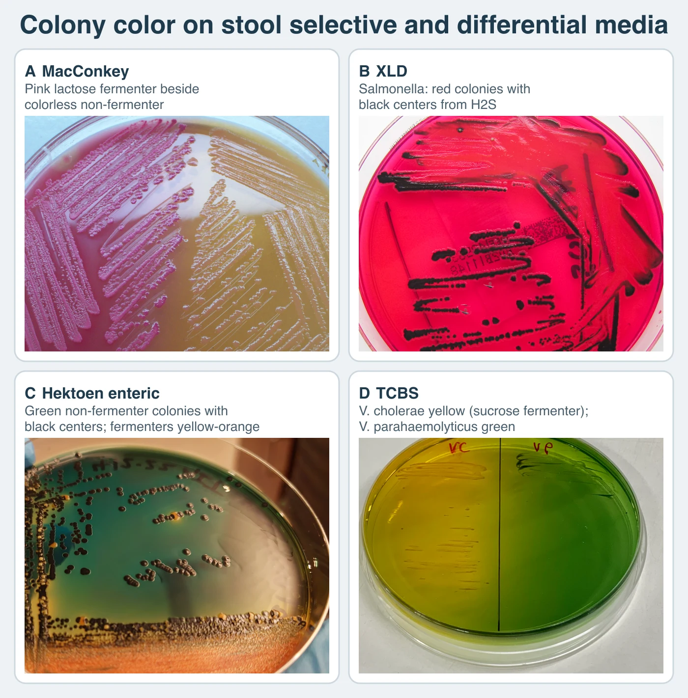
Most laboratories that still culture stool recover Salmonella, Shigella, and Campylobacter routinely. Also test the same specimen for Shiga toxin-producing Escherichia coli (STEC) by toxin immunoassay or stx NAAT, with concurrent culture for O157 on SMAC, CT-SMAC, or a chromogenic O157 agar. Sorbitol screening alone is not a substitute, because it misses the non-O157 serogroups that now cause much of the disease. Recovery of Vibrio and Yersinia in the United States is usually a special request that requires added media or incubation conditions, so the laboratory needs to know the clinical suspicion in advance.2,3,6
Targeted stool pathogen workup
Scroll sideways to see all columns
| Organism or group | Preferred detection | Key confirmatory features |
|---|---|---|
| Salmonella, Shigella | Culture on EMB/MacConkey plus XLD/HE, or multiplex NAAT | Typical nontyphoidal Salmonella: H2S-positive, motile, lysine-decarboxylase positive, serotyped by O and H antigens. S. Typhi is weakly H2S-positive and expresses the Vi capsular antigen; S. Paratyphi A is H2S-negative. Shigella: non-motile, H2S-negative, serogrouped by O antigen |
| STEC, including O157:H7 | Shiga toxin immunoassay or stx NAAT, performed together with O157 culture | stx1 and stx2 genes or toxin detection; O157 strains are classically sorbitol-negative and MUG-negative. Non-O157 STEC ferment sorbitol and are missed by that screen alone |
| Yersinia enterocolitica | CIN agar at 25 °C, or NAAT | Grows on routine media as well. Cold enrichment at 4 °C can improve recovery from mixed or convalescent specimens but delays isolation and is not used for acute diagnosis |
| Campylobacter jejuni and C. coli | Selective microaerophilic media (mCCDA, Campy-CVA) at 42 °C, or NAAT | Gray, flat, spreading colonies; oxidase-positive; curved, S-shaped, or gull-wing gram-negative rods. Stool antigen immunoassay is not a standalone method: a positive result needs culture or NAAT confirmation, and a negative result does not exclude infection |
| Vibrio, Aeromonas, Plesiomonas | TCBS for Vibrio; blood agar for Aeromonas; routine enteric media for Plesiomonas | Oxidase from blood agar, then identification as described below |
| Clostridioides difficile | Multistep algorithm combining glutamate dehydrogenase (GDH) antigen, toxin A/B immunoassay, and NAAT | Testing criteria and algorithm choices appear with the anaerobes below |
| VRE carriage | Vancomycin-containing selective agar or chromogenic VRE agar, with NAAT to separate vanA from vanB | Surveillance and infection-control indication, outside the diarrhea workup |
An E. coli serotype designation combines the O somatic lipopolysaccharide antigen with the H flagellar antigen, and for some strains the K capsular antigen. O157:H7 is the best-known example. It does not ferment sorbitol and lacks the beta-glucuronidase activity that produces fluorescence in the 4-methylumbelliferyl-beta-D-glucuronide (MUG) test, and both reactions are atypical for the species. Screen a sorbitol-negative colony with O157 latex or antiserum, identify it as E. coli, and test it for Shiga toxin or stx genes, because agglutination alone does not establish that an isolate produces toxin. H7 flagellar typing is not performed by slide agglutination in the clinical laboratory. Motile O157 isolates are sent to a public health laboratory, where H type is determined by tube agglutination after motility enhancement or, more often now, predicted from the whole-genome sequence. Serogroups O26, O45, O103, O111, O121, and O145 account for a substantial share of United States STEC disease, and most ferment sorbitol normally. Public health laboratories also perform their serogrouping.6,7
Enterobacterales
The Enterobacterales are facultatively anaerobic, non-spore-forming gram-negative bacilli that ferment glucose with acid production, reduce nitrate to nitrite, and are typically oxidase-negative. Clinically important genera include Citrobacter, Cronobacter, Enterobacter, Escherichia, Hafnia, Klebsiella, Morganella, Proteus, Providencia, Salmonella, Serratia, Shigella, and Yersinia. Plesiomonas shigelloides belongs to the order and is its only oxidase-positive member of clinical importance. Nitrate-negative isolates are rare among the organisms a clinical laboratory reports.1,8
Endotoxin, the lipopolysaccharide of the outer membrane, drives much of the morbidity of gram-negative infection: fever, hypotension, granulocytosis followed by consumption, thrombocytopenia, disseminated intravascular coagulation, and complement activation that can end in septic shock. Individual organisms add their own virulence factors. The K1 capsule appears in E. coli strains causing neonatal meningitis, K. pneumoniae carries an antiphagocytic capsule, and the Vi capsular antigen of S. Typhi reduces complement deposition and neutrophil phagocytosis. Fimbrial adhesins anchor the organism to the mucosa.1,9
Diarrheagenic E. coli are grouped by pathotype: Shiga toxin-producing (STEC), enterotoxigenic (ETEC), enteroinvasive (EIEC), enteropathogenic (EPEC), enteroaggregative (EAEC), and possibly diffusely adherent (DAEC). Enterohemorrhagic E. coli names only the STEC strains that cause human illness, particularly bloody diarrhea and hemolytic uremic syndrome. Hybrid strains carrying virulence factors from more than one pathotype occur. In enterotoxigenic disease, heat-labile toxin activates adenylate cyclase and raises cyclic AMP. Heat-stable toxin activates guanylate cyclase. Both toxins produce secretory watery diarrhea.10
Colony morphology narrows the possibilities but does not identify an isolate. Useful clues include swarming Proteus on blood agar, mucoid and lactose-positive Klebsiella, H2S-producing Salmonella on HE or XLD, indole-positive E. coli, and the occasional red-pigmented S. marcescens. Prodigiosin is often absent at 35 to 37 °C, and most clinical isolates never show it. Matrix-assisted laser desorption/ionization time-of-flight (MALDI-TOF) mass spectrometry identifies most Enterobacterales within minutes, but commercial databases do not reliably separate Shigella species from E. coli, including enteroinvasive strains. Report such isolates as E. coli/Shigella until motility, lysine decarboxylase, acetate or mucate reactions and serology, or an equivalent public-health algorithm, finish the identification.1,11
Biochemical differentiation of representative organisms, where + is at least 90% positive, − is at least 90% negative, V is variable, and H2S is read on triple sugar iron (TSI) agar:
Scroll sideways to see all columns
| Organism | Indole | H2S (TSI) | Urease | Motility | Lactose |
|---|---|---|---|---|---|
| E. coli | + | − | − | + | + |
| Klebsiella pneumoniae/oxytoca | −/+ | − | + | − | + |
| Proteus mirabilis | − | + | + | + (swarms) | − |
| Proteus vulgaris | + | + | + | + (swarms) | − |
| Shigella spp. | V | − | − | − | − (S. sonnei is often positive in the o-nitrophenyl-β-D-galactopyranoside (ONPG) β-galactosidase test, with delayed lactose fermentation) |
| Citrobacter freundii | − | + | V | + | V (delayed) |
| Yersinia enterocolitica | V | − | + | + at 25 °C, − at 37 °C | − |
| Enterobacter cloacae | − | − | V | + | + |
| Serratia marcescens | − | − | V | + | − |
| Morganella morganii | + | − | + | + | − |
| Providencia spp. | + | − | V | + | − |
| Salmonella (typical serotypes) | − | + | − | + | − |
Susceptibility in this order is unpredictable, so test isolates whenever therapy is planned. Uncomplicated Salmonella enteritis is an exception, because antimicrobial therapy does not shorten the diarrhea and can prolong carriage. Mixed-flora infections are another exception, because a single-isolate result may not describe the infection. Basal chromosomal AmpC production predicts intrinsic resistance to ampicillin, amoxicillin-clavulanate, and first-generation cephalosporins in the Enterobacter cloacae complex, Klebsiella aerogenes (formerly Enterobacter aerogenes), the Citrobacter freundii complex, S. marcescens, Providencia species, M. morganii, and Hafnia alvei, and to ampicillin-sulbactam and cephamycins in all of them except Providencia and M. morganii. The risk of clinically important AmpC induction during therapy differs across that list. It is moderate in the E. cloacae complex, K. aerogenes, C. freundii, and H. alvei. M. morganii, Providencia, and S. marcescens have a low risk, and their susceptibility results are reported as tested. Aeromonas and Pseudomonas aeruginosa show a similar basal pattern without belonging to the Enterobacterales.12,13
Extended-spectrum beta-lactamases (ESBLs) and acquired AmpC enzymes are distinct phenotypes with different resistance patterns. An ESBL inactivates most penicillins, extended-spectrum cephalosporins, and aztreonam. It does not inactivate cephamycins or carbapenems, so cephamycin resistance suggests AmpC. Acquired AmpC inactivates cephamycins as well as extended-spectrum cephalosporins, and carbapenems usually remain active unless porin loss accompanies the enzyme. ESBLs are most common in E. coli, K. pneumoniae, and K. oxytoca, where a ceftriaxone minimum inhibitory concentration of 4 µg/mL or greater serves as the practical United States surrogate. Routine phenotypic confirmation is not required when current Clinical and Laboratory Standards Institute (CLSI) breakpoints are used for reporting. Plasmid-mediated AmpC remains less common and is seen mainly in E. coli and K. pneumoniae.12,13
Carbapenem-resistant Enterobacterales (CRE) are resistant to at least one carbapenem or produce a carbapenemase, and those two definitions do not select the same isolates. In many CRE, resistance comes from an ESBL or AmpC enzyme combined with porin loss, with no carbapenemase present. Proteus, Morganella, and Providencia can show intrinsically elevated imipenem minimum inhibitory concentrations that do not make them CRE. Test a carbapenem-nonsusceptible isolate for carbapenemase production and, when indicated, for KPC, NDM, VIM, IMP, and OXA-48-like genes, and notify the clinical and infection-prevention teams immediately. The enzyme family matters for both treatment and containment. KPC remains important, but NDM-producing CRE rose substantially in the United States between 2019 and 2023. Method selection, breakpoints, and the phenotypic and genotypic detection assays themselves are covered in Antimicrobial Susceptibility Testing and Resistance Mechanisms.12-14
Curved and oxidase-positive enteric organisms
Scroll sideways to see all columns
| Feature | Campylobacter jejuni/coli | Helicobacter pylori | Vibrio cholerae | V. parahaemolyticus and V. vulnificus | Aeromonas spp. | Plesiomonas shigelloides |
|---|---|---|---|---|---|---|
| Morphology | Small curved, S-shaped, or gull-wing; microaerophilic | Spiral, microaerophilic | Curved to comma-shaped | Curved to comma-shaped | Straight rod | Straight rod |
| Oxidase | + | + | + | + | + | + |
| NaCl requirement | No | No | No | Yes (halophilic) | No | No |
| Isolation temperature | 42 °C for the thermophilic species | 35-37 °C, humid microaerobic | 35-37 °C | 35-37 °C | 35-37 °C | 35-37 °C (minimum about 8 °C) |
| Identification approach | Hippurate hydrolysis positive in C. jejuni; C. coli is typically negative. Nalidixic-acid and cephalothin disks are obsolete for identification. MALDI-TOF or species-specific polymerase chain reaction (PCR) confirms | Urease strongly positive, the basis of the urea breath test | Sucrose-fermenting, yellow on TCBS | V. parahaemolyticus is sucrose-negative and green on TCBS. Most V. vulnificus are green, a minority yellow, so colony color does not exclude the species. The Kanagawa test is historical and not used clinically | Oxidase-positive, glucose-fermenting straight rods growing without added NaCl; often beta-hemolytic, though A. caviae may be delayed or nonhemolytic. Identify by MALDI-TOF and report a complex when the database cannot separate A. dhakensis from A. hydrophila | Oxidase-positive member of the Enterobacterales; ferments myo-inositol; lysine, ornithine, and arginine all positive; no gas from glucose. Lactose is negative or delayed, so colony color on MacConkey does not rule it out |
| Principal disease | Enteritis, and a recognized antecedent of Guillain-Barré syndrome and reactive arthritis | Gastritis, peptic ulcer disease, gastric adenocarcinoma, mucosa-associated lymphoid tissue (MALT) lymphoma | Epidemic cholera from toxigenic O1 and O139 strains; non-O1/non-O139 strains cause sporadic gastroenteritis, wound infection, and septicemia | V. parahaemolyticus: gastroenteritis after raw or undercooked shellfish. V. vulnificus: rapidly progressive wound infection after seawater exposure, and primary septicemia after raw oysters in liver disease or immunocompromise, fatal in roughly 1 of 5 cases | Watery or dysenteric gastroenteritis with incompletely defined pathogenesis; wound infection after freshwater or soil exposure; septicemia, especially with malignancy or hepatobiliary disease | Gastroenteritis after shellfish ingestion or travel in tropical and subtropical regions; rare meningitis and septicemia |
| Susceptibility notes | Most infections are self-limited. Ciprofloxacin resistance is established in the United States, roughly 28% to 40% by species and year, so ciprofloxacin activity cannot be assumed. Test ciprofloxacin and a macrolide by a Campylobacter-validated method | Clarithromycin and levofloxacin are used only when susceptibility is confirmed by culture or a validated molecular assay. CLSI does not recommend phenotypic metronidazole testing, because results do not predict response | Testing is not routine, because rehydration is the primary treatment. When testing is performed, CLSI M45 criteria apply; O1 and O139 isolates must be submitted to public health | Severe V. vulnificus infection needs immediate treatment, so report an isolate promptly under your laboratory's notification procedure. Testing follows CLSI M45 and is uncommon in routine stool workup | Inducible chromosomal penicillinases and AmpC enzymes make ampicillin and first-generation cephalosporins unreportable. The chromosomal metallo-carbapenemase CphA is common in A. hydrophila, A. veronii, and A. dhakensis, usually absent in A. caviae, and easily missed at standard inoculum | Chromosomal beta-lactamase usually confers ampicillin resistance. Interpret with CLSI M100 Enterobacterales breakpoints. Fluoroquinolones and carbapenems are usually active |
An oxidase-positive, spiral gram-negative rod that hydrolyzes sodium hippurate may be reported as C. jejuni without further testing, though some strains hydrolyze weakly or not at all and then require MALDI-TOF or species-specific PCR. Biochemical and disk-based schemes, including the nalidixic-acid and cephalothin disks that older references describe, are no longer accepted for species identification. Their accuracy is poor, and fluoroquinolone resistance is now common enough that a nalidixic-acid result has no identification value. Incubate primary selective agar 48 to 72 hours at 41 to 42.5 °C in a microaerophilic atmosphere of roughly 5% O2, 10% CO2, and 85% N2. That temperature favors C. jejuni and C. coli. It misses species such as C. upsaliensis and C. fetus, which grow better at 35 to 37 °C, often with hydrogen enrichment. C. fetus is a bloodstream organism and is not thermophilic. It causes bacteremia in immunocompromised, pregnant, or elderly patients and is recovered from blood. Selective plates incubated at 42 °C miss it.15-17
H. pylori testing depends on whether endoscopy is otherwise indicated. When it is not, active infection is established with a urea breath test or a monoclonal stool antigen immunoassay, both preferred over serology, which cannot separate current from past infection. When endoscopy is performed, the options are biopsy urease, histology, and, where susceptibility is needed, culture or gastric NAAT. Clarithromycin and levofloxacin resistance is common in the United States, so clinicians often request a culture-based or molecular susceptibility result for these agents, whether or not the patient has been treated before. When culture is pursued, incubate it at 35 to 37 °C for at least 10 days and up to 14 in a humid microaerobic atmosphere of about 5% O2 and 5% to 10% CO2 with the balance nitrogen. Hydrogen improves recovery. A candle jar or a CO2-only incubator will not grow the organism.18,19
V. cholerae and V. mimicus grow without added NaCl. The other medically important species need salt. Perform oxidase from blood agar colonies, never from TCBS or MacConkey colonies, because both media can give an incorrect result. The 0.5% sodium deoxycholate string test, usually positive in Vibrio and negative in Aeromonas, is a historical screen. Current public-health workflows use MALDI-TOF, species-specific PCR, or whole-genome sequencing. A classic biochemical scheme still separates the species using growth in nutrient broth with 0% or 1% NaCl, oxidase, nitrate reduction, myo-inositol fermentation, arginine dihydrolase, lysine and ornithine decarboxylase, and sucrose on TCBS, and it remains a useful backup when a rapid method is unavailable or inconclusive:1,20
Scroll sideways to see all columns
| Test | V. cholerae | V. mimicus | V. parahaemolyticus | V. vulnificus | V. alginolyticus |
|---|---|---|---|---|---|
| Growth in 0% NaCl | + | + | − | − | − |
| Growth in 1% NaCl | + | + | + | + | + |
| Oxidase | + | + | + | + | + |
| Nitrate to nitrite | + | + | + | + | + |
| myo-Inositol | − | − | − | − | − |
| Arginine dihydrolase | − | − | − | − | − |
| Lysine decarboxylase | + | + | + | + | + |
| Ornithine decarboxylase | + | + | + | V | V |
| Sucrose (TCBS) | + (yellow) | − (green) | − (green) | Usually − (green); variable | + (yellow) |
Most V. vulnificus isolates are sucrose-negative and green on TCBS, but a minority ferment sucrose and appear yellow, so colony color never excludes the species. Use MALDI-TOF, species-specific PCR, or whole-genome sequencing to resolve an inconclusive biochemical result. Sequencing is performed by public health laboratories.20,21
Clostridioides difficile and gastrointestinal anaerobes
Anaerobes dominate the flora of the gastrointestinal tract and appear routinely in mixed wound and abscess cultures after mucosal or tissue disruption. For this reason, stool is not an acceptable specimen for general anaerobic culture. An anaerobe recovered from feces does not indicate infection. Two targeted exceptions exist. Toxigenic C. difficile culture is a reference procedure, and public health laboratories test feces for C. botulinum and for C. perfringens foodborne enterotoxin. Perform general anaerobic culture on tissue, aspirates, and other properly collected specimens.2,22
For those specimens, obligate anaerobes are defined operationally: they form no colonies on solid media incubated in air or in 10% CO2 in air. Oxygen tolerance varies within the category. Some strict species are inhibited near 0.5% O2. Many clinically important moderate anaerobes, including the Bacteroides fragilis group, tolerate about 2% to 8%. Aerotolerant anaerobes may produce scant growth in room air with CO2 but do not use oxygen for energy. Plate specimens to an enriched non-selective medium such as Brucella, CDC, or Columbia agar with sheep blood, vitamin K, and hemin, along with selective and differential agars including kanamycin-vancomycin laked blood, Bacteroides bile esculin, phenylethyl alcohol, and colistin-nalidixic acid, plus a broth. Incubate at 35 to 37 °C in an oxygen-free atmosphere that typically contains 5% to 10% H2 and 5% to 10% CO2 with the balance nitrogen, verified by a chemical or biologic indicator. Keep jars, boxes, and pouches closed for at least 48 hours, because air reaching a culture during logarithmic growth kills slower-growing species. Plates inside an anaerobic chamber may be examined at 24 hours. Keep primary plates 5 to 7 days when slow growers are a concern.22
C. difficile testing starts with diagnostic stewardship, because asymptomatic carriage is common and no single assay distinguishes carriage from disease. Testing is limited to unexplained new-onset diarrhea, meaning at least three unformed stools in 24 hours, from a patient who has not received a laxative in the previous 48 hours. Formed stool is rejected unless the physician documents ileus or possible toxic megacolon. Infants 12 months of age or younger are not tested, and children between 1 and 2 years are not tested routinely until other infectious and noninfectious causes have been excluded. Where a laboratory has no preagreed submission criteria, use a multistep algorithm that includes a toxin assay: GDH with toxin, GDH with toxin arbitrated by NAAT, or NAAT with toxin. Where such criteria exist, NAAT alone or a multistep algorithm is acceptable. GDH is produced by toxigenic and non-toxigenic strains alike, so a GDH-positive, toxin-negative result is inconclusive, and a NAAT-positive, toxin-negative result often reflects carriage. Toxigenic culture and cell-culture cytotoxicity neutralization remain reference methods. Enterotoxin TcdA and cytotoxin TcdB damage the colonic epithelium and produce antibiotic-associated diarrhea and pseudomembranous colitis. The 2021 Infectious Diseases Society of America (IDSA)/Society for Healthcare Epidemiology of America (SHEA) focused update revised treatment and left this diagnostic approach unchanged.2,4,24
C. perfringens is common fecal flora, so routine enteric culture does not look for it, and public-health enterotoxin testing confirms foodborne illness. Recovered from tissue or an aspirate in a compatible clinical setting, it forms large gray colonies with a double zone of beta-hemolysis, and the cells are boxcar-shaped gram-positive rods that are usually nonmotile, lecithinase-positive, lipase-negative, and reverse CAMP-positive (synergistic arrowhead hemolysis with a group B Streptococcus streak). Isolation from a wound without clinical myonecrosis does not diagnose gas gangrene.2,22
Anaerobic susceptibility testing is not performed on stool isolates. It is considered when a clinically significant anaerobe comes from a normally sterile site, grows in pure culture, follows a treatment failure, or will be treated for a prolonged course, and it matters most for organisms with unpredictable resistance, especially Bacteroides and Parabacteroides, along with selected Prevotella, Fusobacterium, and Clostridium isolates. The CLSI reference method is agar dilution on supplemented Brucella blood agar. Broth microdilution in supplemented Brucella broth with hemin, vitamin K1, and 5% lysed horse blood is an alternative for Bacteroides and Parabacteroides with selected agents. Gradient minimum inhibitory concentration tests are widely used for individual isolates when validated against the reference method. CLSI does not provide a routine anaerobic disk-diffusion method. The European Committee on Antimicrobial Susceptibility Testing (EUCAST) publishes a separate standardized disk-diffusion method for selected rapidly growing anaerobes. Its media, breakpoints, and categories are not interchangeable with the CLSI methods taught here.26 Under CLSI, breakpoints come from the current M100 anaerobic tables. Routine susceptibility testing of C. difficile from stool is not indicated outside public-health surveillance or an extraintestinal isolate.13,25 At the bench, check each stool request against the specimen, the collection day, and the organisms the requested method detects.
Practice
References
- Tille PM. Bailey & Scott's Diagnostic Microbiology. 16th ed. Elsevier; 2025. Back to text
- Miller JM, Binnicker MJ, Campbell S, et al. Guide to utilization of the microbiology laboratory for diagnosis of infectious diseases: 2024 update by the Infectious Diseases Society of America and the American Society for Microbiology. Clin Infect Dis. Published online March 5, 2024. doi:10.1093/cid/ciae104 Back to text
- Shane AL, Mody RK, Crump JA, et al. 2017 Infectious Diseases Society of America clinical practice guidelines for the diagnosis and management of infectious diarrhea. Clin Infect Dis. 2017;65(12):e45-e80. doi:10.1093/cid/cix669 Back to text
- McDonald LC, Gerding DN, Johnson S, et al. Clinical practice guidelines for Clostridium difficile infection in adults and children: 2017 update by the Infectious Diseases Society of America (IDSA) and Society for Healthcare Epidemiology of America (SHEA). Clin Infect Dis. 2018;66(7):e1-e48. doi:10.1093/cid/cix1085 Back to text
- Bauer TM, Lalvani A, Fehrenbach J, et al. Derivation and validation of guidelines for stool cultures for enteropathogenic bacteria other than Clostridium difficile in hospitalized adults. JAMA. 2001;285(3):313-319. doi:10.1001/jama.285.3.313 Back to text
- Gould LH, Bopp C, Strockbine N, et al. Recommendations for diagnosis of Shiga toxin-producing Escherichia coli infections by clinical laboratories. MMWR Recomm Rep. 2009;58(RR-12):1-14. Accessed August 31, 2026. https://www.cdc.gov/mmwr/preview/mmwrhtml/rr5812a1.htm Back to text
- Association of Public Health Laboratories. Identification Guide: Isolation and Identification of Shiga Toxin-Producing Escherichia coli from Culture-Independent Diagnostic Test Positive Specimens. APHL; April 2025. Accessed August 31, 2026. https://www.aphl.org/aboutAPHL/publications/Documents/FS-STEC-CIDT-Guide.pdf Back to text
- Janda JM, Abbott SL. The changing face of the family Enterobacteriaceae (order: Enterobacterales): new members, taxonomic issues, geographic expansion, and new diseases and disease syndromes. Clin Microbiol Rev. 2021;34(2):e00174-20. doi:10.1128/CMR.00174-20 Back to text
- Hiyoshi H, Wangdi T, Lock G, et al. The Vi capsular polysaccharide of Salmonella Typhi promotes macrophage phagocytosis by binding the human C-type lectin DC-SIGN. mBio. 2022;13(5):e02733-22. doi:10.1128/mbio.02733-22 Back to text
- Centers for Disease Control and Prevention. Technical information: E. coli infection. Updated May 14, 2024. Accessed August 31, 2026. https://www.cdc.gov/ecoli/php/technical-info/index.html Back to text
- Association of Public Health Laboratories. Identification Guide: Isolation and Identification of Shigella species and Enteroinvasive Escherichia coli from Culture-Independent Diagnostic Test Positive Specimens. APHL; July 2025. Accessed August 31, 2026. https://www.aphl.org/aboutAPHL/publications/Documents/FS-Shigella-EIEC-CIDT-Guide.pdf Back to text
- Infectious Diseases Society of America. Guidance on the Treatment of Antimicrobial Resistant Gram-Negative Infections. IDSA; 2026. Accessed August 31, 2026. https://www.idsociety.org/practice-guideline/amr-guidance/ Back to text
- CLSI. Performance Standards for Antimicrobial Susceptibility Testing. 36th ed. CLSI supplement M100. Clinical and Laboratory Standards Institute; 2026. Back to text
- Centers for Disease Control and Prevention. Carbapenem-resistant Enterobacterales (CRE) infection control. Updated December 17, 2025. Accessed August 31, 2026. https://www.cdc.gov/cre/hcp/infection-control/index.html Back to text
- Association of Public Health Laboratories. Campylobacter Isolation and Characterization from Clinical Specimens: Guidance for Public Health Laboratories. APHL; September 2023. Accessed August 31, 2026. https://www.aphl.org/aboutAPHL/publications/Documents/FS-Campylobacter-Diagnosis-Recommendations.pdf Back to text
- CLSI. Methods for Antimicrobial Dilution and Disk Susceptibility Testing of Infrequently Isolated or Fastidious Bacteria. 3rd ed. CLSI guideline M45. Clinical and Laboratory Standards Institute; 2016. Back to text
- Centers for Disease Control and Prevention. Clinical overview of Campylobacter. Updated August 12, 2026. Accessed September 24, 2026. https://www.cdc.gov/campylobacter/hcp/clinical-overview/index.html Back to text
- Chey WD, Howden CW, Moss SF, et al. ACG clinical guideline: treatment of Helicobacter pylori infection. Am J Gastroenterol. 2024;119(9):1730-1753. doi:10.14309/ajg.0000000000002968 Back to text
- Schuetz AN, Cornish NE, Doern CD, et al. Testing for Helicobacter pylori in an era of antimicrobial resistance. J Clin Microbiol. 2024;62(2):e00732-23. doi:10.1128/jcm.00732-23 Back to text
- Association of Public Health Laboratories. Identification Guide: Isolation and Identification of Vibrio Species from Culture-Independent Diagnostic Test Positive Specimens. APHL; January 2026. Accessed August 31, 2026. https://www.aphl.org/aboutAPHL/publications/Documents/FS-Vibrio-CIDT-Guide.pdf Back to text
- Centers for Disease Control and Prevention. Clinical overview of vibriosis. Updated May 13, 2024. Accessed August 31, 2026. https://www.cdc.gov/vibrio/hcp/clinical-overview/index.html Back to text
- Nagy E, Boyanova L, Justesen US. How to isolate, identify and determine antimicrobial susceptibility of anaerobic bacteria in routine laboratories. Clin Microbiol Infect. 2018;24(11):1139-1148. doi:10.1016/j.cmi.2018.02.008 Back to text
- George WL, Sutter VL, Citron D, Finegold SM. Selective and differential medium for isolation of Clostridium difficile. J Clin Microbiol. 1979;9(2):214-219. doi:10.1128/jcm.9.2.214-219.1979 Back to text
- Johnson S, Lavergne V, Skinner AM, et al. Clinical practice guideline by the Infectious Diseases Society of America (IDSA) and Society for Healthcare Epidemiology of America (SHEA): 2021 focused update guidelines on management of Clostridioides difficile infection in adults. Clin Infect Dis. 2021;73(5):e1029-e1044. doi:10.1093/cid/ciab549 Back to text
- CLSI. Methods for Antimicrobial Susceptibility Testing of Anaerobic Bacteria. 9th ed. CLSI standard M11. Clinical and Laboratory Standards Institute; 2018. Reaffirmed April 2025. Back to text
- European Committee on Antimicrobial Susceptibility Testing. Disk diffusion and quality control: EUCAST disk diffusion method for antimicrobial susceptibility testing of anaerobic bacteria, version 3.0, June 2026. Accessed September 13, 2026. https://www.eucast.org/bacteria/methodology-and-instructions/disk-diffusion-and-quality-control/ Back to text