Hyphae invading viable tissue support mold infection but seldom reveal which genus is responsible. Culture, matrix-assisted laser desorption/ionization time-of-flight (MALDI-TOF) mass spectrometry, or sequencing can identify an organism, but recovery from a nonsterile site does not prove invasion. Interpretation must account for the specimen, direct examination, histopathology, culture, host factors, and radiologic or operative findings.1
Specimen interpretation and mold safety
Submit tissue for both microbiology and histopathology when invasive mold disease is suspected. Place the microbiology portion in a sterile container without formalin and the pathology portion in formalin. Direct potassium hydroxide (KOH)-calcofluor examination can show fungal elements within minutes. Hematoxylin and eosin (H&E) staining demonstrates tissue injury and host response. Gomori methenamine silver (GMS) and periodic acid-Schiff (PAS) stains improve fungal contrast. A negative direct examination or culture does not exclude infection, particularly after antifungal exposure or when little specimen was collected.
Scroll sideways to see all columns
| Finding | What it supports | What it does not establish |
|---|---|---|
| Hyphae invading viable tissue or vessels | Invasive mold disease | Genus or species from branch angle, width, or septation alone |
| Mold from a normally sterile biopsy, with matching histology | The recovered mold is likely etiologic | That a negative culture excludes the mold seen in tissue |
| Mold from sputum, bronchoalveolar lavage (BAL), sinus material, skin, or another nonsterile site | Exposure, colonization, contamination, or infection, depending on the setting | Invasion by itself |
| Repeated recovery of the same mold with compatible direct findings and disease | Greater likelihood of clinical significance | A universal rule that separates colonization from infection |
| One environmental mold colony without direct evidence | Possible contamination, especially from a nonsterile specimen | That the isolate can always be dismissed |
Handle every filamentous or fuzzy colony in a certified Class II biological safety cabinet, including molds first noticed on bacteriology media. Keep plates closed and sealed until work begins in the cabinet. Never smell a culture. Do not prepare open-bench tape mounts, slide cultures, or other procedures that can aerosolize conidia. If an unidentified mold may be a thermally dimorphic pathogen or another high-risk organism, stop routine manipulation and follow your laboratory's containment and referral procedure.2
Dermatophytes
Dermatophytes infect keratinized skin, hair, and nails. The traditional clinical genera Trichophyton, Microsporum, and Epidermophyton remain useful, but molecular taxonomy also recognizes Nannizzia, Lophophyton, Paraphyton, and Arthroderma. Some organisms formerly reported as Microsporum, including M. gypseum, now belong to Nannizzia. Report the name supported by your laboratory's validated database and include a familiar former name when it prevents confusion.3,4
Tinea names describe the infected site and do not indicate the causative genus: capitis involves the scalp, barbae the beard area, corporis glabrous skin, cruris the groin, pedis the foot, manuum the hand, and unguium the nail. Trichophyton species may infect all three keratinized sites. Microsporum species primarily infect hair and skin, and Epidermophyton floccosum infects skin and nails but not hair.
Collect skin scale from the active border, affected hair stubs with scalp scale, and nail plate with subungual debris from the advancing diseased area. KOH with or without a fluorescent brightener demonstrates hyaline septate hyphae and arthroconidia but does not identify a species. In hair, arthroconidia within the shaft indicate endothrix invasion. A sheath around the shaft indicates ectothrix invasion. Some ectothrix infections caused by M. canis, M. audouinii, or M. ferrugineum fluoresce blue-green to yellow-green under a Wood lamp. Fluorescence is inconsistent, and a negative examination does not exclude tinea capitis. T. tonsurans produces endothrix infection and does not fluoresce.
Inoculate a nonselective fungal medium and, when appropriate, a medium containing antibacterial agents and cycloheximide. Cycloheximide suppresses many environmental molds but can also inhibit clinically important nondermatophyte fungi, so never use it as the only medium. Dermatophyte test medium color change is presumptive because nondermatophyte molds can also alkalinize the medium. Direct microscopy and culture are complementary. Either can be negative after treatment or with a poor specimen.
Scroll sideways to see all columns
| Taxon or reporting group | Useful presumptive clues in culture | Important limit |
|---|---|---|
| Microsporum canis | Numerous rough, thick-walled, spindle-shaped macroconidia, often with more than six septa; few microconidia | Pigment and Wood lamp fluorescence vary |
| M. audouinii and M. ferrugineum | Often poorly sporulating; distorted or pectinate hyphae and terminal chlamydoconidia may occur | Phenotype may be insufficient for species identification |
| Nannizzia gypsea complex, formerly M. gypseum complex | Numerous thin-walled, rough, symmetric ellipsoidal macroconidia with relatively few septa | Members of the complex overlap morphologically |
| Trichophyton rubrum complex | Small pyriform microconidia along the sides of hyphae; pencil-shaped macroconidia are uncommon; a red reverse may develop on a pigment-promoting medium | Red pigment, urease negativity, and a negative hair-perforation test are variable presumptive traits |
| T. mentagrophytes/T. interdigitale complex | Round microconidia in clusters, spiral hyphae, and occasional cigar-shaped macroconidia; many isolates are urease positive | Urease, hair perforation, colony texture, and internal transcribed spacer (ITS) sequencing alone may not resolve closely related members |
| T. tonsurans | Variable microconidia, including elongated and balloon forms, along irregular hyphae; macroconidia are uncommon | Colony color and texture vary widely |
| T. verrucosum | Slow growth, few conidia, and chains of chlamydoconidia; cattle exposure may support the identification | Nutritional tests and morphology are slow and only presumptive |
| Epidermophyton floccosum | Smooth, club-shaped macroconidia in clusters; microconidia absent | Old or pleomorphic cultures may lose typical sporulation |
Morphology is useful for a provisional genus, species complex, or familiar species pattern. Do not report a species from morphology when structures are absent or overlapping. This is especially important for T. indotineae, which routine phenotypic methods commonly report as T. mentagrophytes or T. interdigitale. The United States has documented T. indotineae, T. mentagrophytes genotype VII, and terbinafine-resistant T. rubrum. Severe, extensive, unusual, or treatment-refractory dermatophytosis warrants referral for validated molecular identification and, when indicated, dermatophyte susceptibility testing. A resistance-associated squalene epoxidase mutation supports terbinafine resistance only when the assay and the association have been validated. Species identification and resistance prediction are separate results.5
Pigmented molds and implantation mycoses
Dematiaceous fungi contain melanin in their cell walls and usually form brown to black colonies. Pigment in tissue may be obvious or subtle. Fontana-Masson staining can support the presence of melanin. Species identification requires culture or sequencing.
Scroll sideways to see all columns
| Syndrome | Defining tissue or specimen finding | Representative fungi and interpretation |
|---|---|---|
| Eumycetoma | Tumefaction, draining sinuses, and fungal grains | Madurella mycetomatis is a major worldwide cause. Scedosporium species can produce pale-grain eumycetoma and are hyaline, so eumycetoma is not limited to pigmented molds. Examine and culture washed grains. Grain color alone does not identify the species. |
| Chromoblastomycosis | Thick-walled brown muriform cells, also called sclerotic bodies, dividing in more than one plane | Common agents include Fonsecaea, Cladophialophora carrionii, and Phialophora verrucosa. Muriform cells define the tissue reaction. Culture or molecular testing identifies the agent. |
| Phaeohyphomycosis | Pigmented septate hyphae, pseudohyphae, yeast-like cells, or a mixture in tissue | Agents include Alternaria, Bipolaris, Curvularia, Exophiala, and Cladophialophora bantiana. The category includes superficial, subcutaneous, cerebral, and disseminated infections. Allergic fungal disease without tissue invasion is not phaeohyphomycosis. |
C. bantiana is neurotropic and may cause cerebral infection in patients without recognized immunodeficiency. Its thermotolerance and long chains of sparsely branched conidia are useful clues, but confirm the identification with a validated molecular or MALDI-TOF database. Pigmented molds are common in the environment. Growth from a nonsterile specimen may represent colonization or contamination. Pigmented hyphae invading tissue establish infection but do not identify the genus.6
Mucorales
Mucormycosis most often occurs with uncontrolled diabetes, especially ketoacidosis, corticosteroid exposure, neutropenia, hematopoietic or solid-organ transplantation, severe trauma or burns, prematurity, or deferoxamine therapy. Clinical forms include rhino-orbital-cerebral, pulmonary, cutaneous, gastrointestinal, and disseminated disease. The major human pathogens belong to Rhizopus, Mucor, Lichtheimia, Rhizomucor, Cunninghamella, Apophysomyces, and Saksenaea.
In tissue, Mucorales usually appear as broad, ribbon-like, irregularly branching hyphae with few septa (pauciseptate). Width and branch angle vary, and folds or fragmentation during tissue processing can create false septa or misleading angles. Report "broad, irregular, pauciseptate hyphae consistent with Mucorales" when that pattern is present. Do not identify Rhizopus, Mucor, or another genus from tissue geometry alone. H&E shows necrosis and angioinvasion. GMS and PAS improve detection of fungal walls.7
Divide tissue before processing. Mince or slice the culture portion with minimal crushing. Do not grind it. Place separate pieces directly onto media without cycloheximide. Culture may remain negative despite visible tissue invasion, so a negative culture does not exclude the infection seen on histopathology. Molecular testing of fresh or formalin-fixed tissue can support identification when culture fails, but performance depends on the assay, fungal burden, fixation, and contamination control.
Scroll sideways to see all columns
| Genus | Presumptive culture morphology |
|---|---|
| Rhizopus | Well-developed nodal rhizoids directly opposite usually unbranched sporangiophores |
| Lichtheimia | Conical apophysis below a pear-shaped sporangium, with sparse internodal rhizoids |
| Mucor | Branched sporangiophores without rhizoids or an apophysis |
| Rhizomucor | Rhizoids with branched sporangiophores, combining features of Rhizopus and Mucor |
| Cunninghamella | Terminal vesicles bearing denticles and one-celled sporangioles |
| Apophysomyces and Saksenaea | Poor or absent sporulation on routine media; molecular identification is often needed |
These structures support genus-level identification only when the culture sporulates typically. ITS or D1/D2 sequencing with a curated database provides stronger identification. Galactomannan and serum (1→3)-β-D-glucan are usually negative in isolated mucormycosis. A negative result does not exclude disease. A positive result does not exclude mucormycosis, because mixed infection and assay cross-reactivity occur.
Aspergillus
Aspergillus causes allergic disease, airway colonization, noninvasive fungus balls, chronic pulmonary disease, tracheobronchitis, and invasive disease. A respiratory isolate therefore has different significance in a patient with an aspergilloma, a lung transplant recipient, a neutropenic patient with nodules, and a patient without compatible disease. Repeated recovery, direct hyphae, positive BAL galactomannan, compatible imaging, and host risk increase the likelihood of infection. Tissue invasion establishes invasive mold disease. Hyaline septate hyphae with acute-angle branching indicate only an Aspergillus-like pattern, because Fusarium, Scedosporium, and other hyaline molds can look the same in tissue.1
An Aspergillus conidial head consists of a conidiophore ending in a vesicle, with phialides directly on the vesicle or on metulae. Colony color, conidial-head arrangement, surface ornamentation, and growth temperature support a provisional section or species complex.
Scroll sideways to see all columns
| Reporting group | Typical culture clues | Laboratory significance |
|---|---|---|
| A. fumigatus complex, section Fumigati | Blue-green to gray-green colony; smooth conidiophore; uniseriate phialides over the upper portion of the vesicle, producing a columnar head | Cryptic species may differ in susceptibility; atypical isolates need molecular confirmation |
| A. flavus complex, section Flavi | Yellow-green colony; rough conidiophore; radiate, uniseriate or biseriate heads; some isolates form sclerotia | Common in sinus, corneal, and invasive disease in some regions |
| A. niger complex, section Nigri | Dark brown to black conidia; large radiate, usually biseriate heads | Black aspergilli overlap morphologically, so report them as a complex unless a validated method resolves them |
| A. terreus complex, section Terrei | Tan to cinnamon colony; compact biseriate heads; accessory conidia may occur along vegetative hyphae | Many isolates have high amphotericin B minimum inhibitory concentrations (MICs), so identify the complex accurately |
Morphology alone does not reliably separate cryptic species. ITS commonly identifies the section or complex. Beta-tubulin or calmodulin sequencing may be needed for species-level resolution. MALDI-TOF can identify an isolate when the extraction method and reference library cover that organism. Report the complex when the validated method cannot support a species.
Other hyaline septate molds
Hyalohyphomycosis describes tissue infection by hyaline septate molds other than the agents assigned a more specific disease name. Histology establishes hyaline mold invasion but rarely distinguishes among these organisms. Culture and a validated identification method are needed.6
Scroll sideways to see all columns
| Mold | Useful clues | Interpretive limits |
|---|---|---|
| Fusarium species complexes | Sickle or canoe-shaped multiseptate macroconidia, phialides with microconidia, and sometimes chlamydoconidia | Macroconidia support the genus only. Adventitious sporulation in tissue helps explain positive blood cultures and metastatic skin lesions in disseminated disease. |
| Scedosporium species | Single oval conidia from annellides; some isolates form synnemata or a sexual state | Respiratory recovery may represent colonization in structurally abnormal lungs. Species-complex members overlap phenotypically. |
| Lomentospora prolificans | Annelloconidia from swollen, flask-shaped annellides | Formerly Scedosporium prolificans. Disseminated infection often yields positive blood cultures and many isolates show resistance to multiple antifungal classes. |
| Purpureocillium lilacinum | Delicate phialides with long tapering necks and chains of lilac conidia | Formerly Paecilomyces lilacinus. Distinguish it from Paecilomyces variotii, which remains in Paecilomyces. |
| Acremonium, Scopulariopsis, Paecilomyces variotii, and other rare molds | Phialides or annellides with distinctive conidial arrangements may suggest a genus | Environmental recovery is common, databases may be incomplete, and clinically important isolates often require sequencing or referral. |
Fusarium is a major cause of keratitis and can cause localized or disseminated disease in severely immunocompromised patients. Scedosporium may cause mycetoma, pulmonary infection, or cerebral infection after near-drowning. Lomentospora prolificans causes localized and disseminated disease but is a separate genus from Scedosporium. Colony color, conidial shape, and blood-culture pattern do not prove infection or identify a species.
Fungal biomarkers and antigen tests
Biomarkers provide mycologic evidence but do not demonstrate an organism in tissue. Their value depends on the population, specimen, assay threshold, prior antifungal exposure, and pretest probability. Correlate an isolated positive result, and often repeat it or confirm it with an orthogonal test.
Scroll sideways to see all columns
| Test | Best-supported use | Major limitations |
|---|---|---|
| Aspergillus galactomannan | Serum or plasma in selected high-risk patients, especially those with hematologic malignancy or hematopoietic stem-cell transplantation; BAL for suspected pulmonary disease | Sensitivity falls during mold-active therapy and may be lower outside neutropenic populations. Cross-reactive fungi, some products or solutions, airway colonization, and specimen contamination can produce positive results. It does not establish tissue invasion or an Aspergillus species. |
| Serum (1→3)-β-D-glucan | Adjunct for several invasive fungal diseases and for Pneumocystis pneumonia | It is not organism-specific. Mucorales and Cryptococcus often produce little detectable analyte. Hemodialysis materials, blood products, glucan-containing gauze, some drugs, bacteremia, and collection contamination can cause false positivity. |
| Cryptococcal capsular antigen | Serum or cerebrospinal fluid (CSF) diagnosis of cryptococcosis | A prozone or postzone effect and rare cross-reactions require method-specific troubleshooting when the result conflicts with the specimen findings. |
| Histoplasma and Blastomyces antigen | Urine, serum, or BAL in the appropriate syndrome and epidemiologic setting | Strong cross-reactivity between endemic fungi limits species attribution. Antigen concentration does not by itself separate localized from disseminated disease. |
Serum galactomannan and β-D-glucan are not screening tests for every mold. Negative results do not exclude invasive aspergillosis, mucormycosis, fusariosis, or another rare mold infection. Consensus thresholds used to classify invasive fungal disease for research are not stand-alone clinical diagnoses.8
Microsporidia
Microsporidia are obligate intracellular, spore-forming eukaryotes closely related to fungi. Their formal taxonomic placement has changed over time. Enterocytozoon bieneusi and Encephalitozoon intestinalis are major causes of chronic diarrhea in people with advanced immunosuppression. Other species can involve the eye, respiratory tract, urinary tract, muscle, or disseminated sites.
Medically important spores usually measure 1 to 4 µm. E. bieneusi spores are about 0.8 to 1.4 µm. Encephalitozoon and several other genera are commonly 1.5 to 4 µm. Modified trichrome with Chromotrope 2R or a quick-hot Gram-chromotrope method stains spores pink-red or violet and may show an equatorial belt. Calcofluor or another fluorescent brightener is a sensitive screen but can also stain yeast and debris. Routine light microscopy does not identify the species. Species-specific polymerase chain reaction (PCR) or broad-range PCR with sequencing is preferred when species identification affects interpretation. Concentrate stool or other fluids according to the validated procedure, use a positive control, and examine enough fields because organism burden may be low.9
Process specimens that may contain microsporidia under biosafety level 2 conditions in a biological safety cabinet, with eye protection. Spores can infect through splash exposure, and laboratory-acquired ocular infection has been reported.9
Pneumocystis
Pneumocystis jirovecii infects humans. P. carinii refers to a rat-associated species. Routine clinical culture is not available. Lower respiratory material is required because oral and upper-airway specimens have lower and less consistent diagnostic yield. BAL generally yields more organisms than induced sputum. Organism burden may be lower in patients without HIV than in those with HIV.
GMS stains the walls of the 4 to 8 µm asci, historically called cysts, but does not reliably stain trophic forms. Giemsa, Wright-Giemsa, Diff-Quik, and related stains show nuclei and trophic forms with less specific morphology. Direct or indirect immunofluorescence with monoclonal antibodies is more sensitive than conventional colorimetric stains. A negative stain does not exclude disease when specimen quality or organism burden is low.
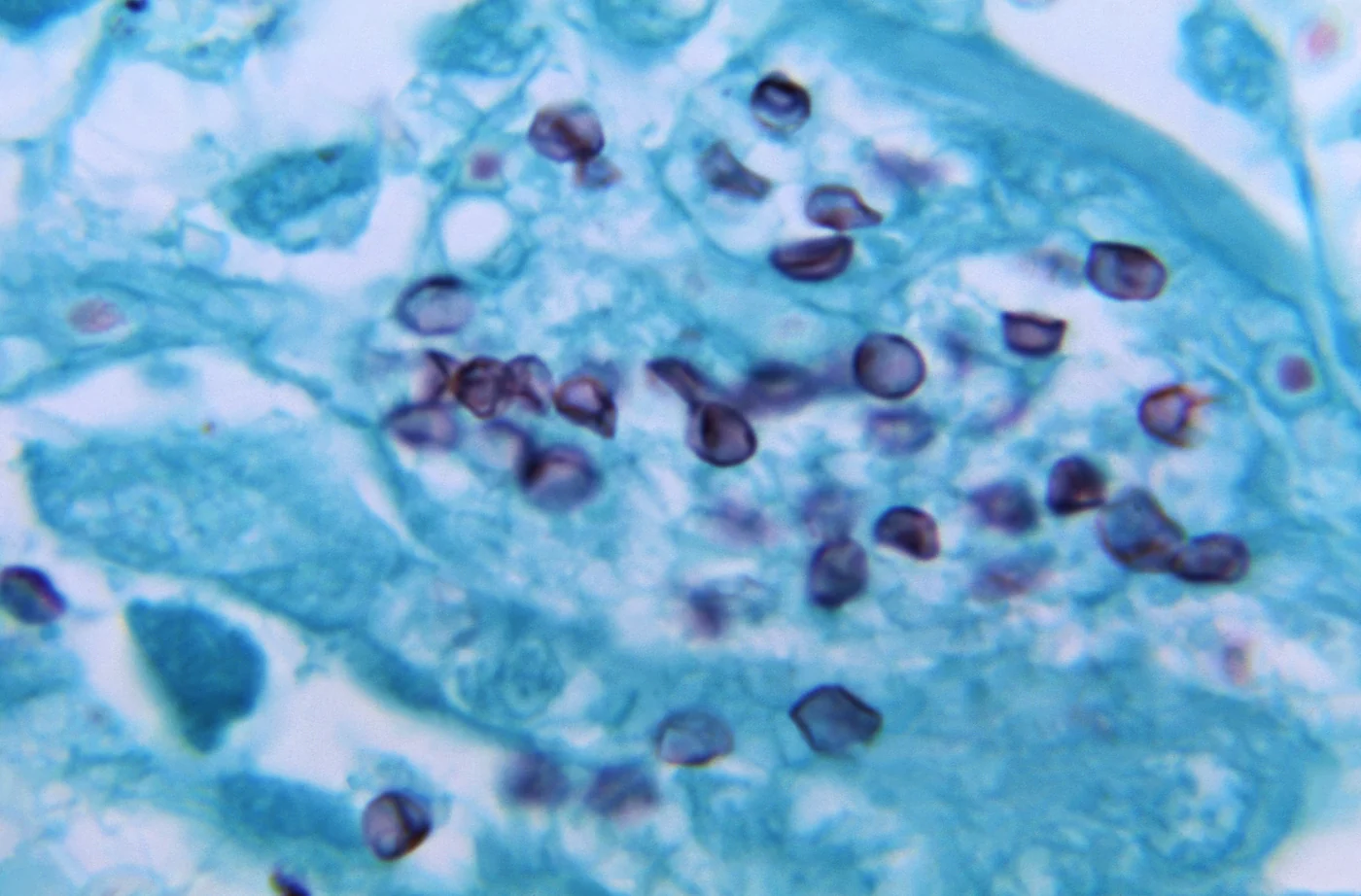
PCR has replaced staining in many laboratories because it is more sensitive. Quantitative PCR is preferable to a qualitative result. No universal cycle-threshold or copy-number cutoff distinguishes pneumonia from colonization across assays and specimen types. A high burden supports disease in a compatible host. A low positive result may represent early infection, resolving infection, or colonization. Interpret it with symptoms, imaging, host risk, specimen type, microscopy, and serum β-D-glucan. β-D-glucan is often elevated, and a low result can make Pneumocystis pneumonia less likely. A positive result is nonspecific and cannot identify Pneumocystis.10
Prototheca
Prototheca species are achlorophyllous green algae that produce yeast-like colonies, so the mycology laboratory often recovers them. P. wickerhamii causes most reported human disease, including cutaneous infection, olecranon bursitis, and disseminated infection. Other species occur less often, and older reports that use P. zopfii may not correspond to current molecular species.
Direct preparations and tissue sections show round to oval, nonbudding cells with internal cleavage into autospores. Mature sporangia have a morula-like or cartwheel appearance and stain with H&E, PAS, GMS, and calcofluor. These structures distinguish Prototheca from most yeasts.3 Morphology and commercial yeast assimilation systems may misidentify the species. MALDI-TOF can identify isolates only if the validated library contains current Prototheca entries. Cytochrome b sequencing provides the strongest species resolution. D1/D2 sequencing is a useful alternative. Interpret growth from a nonsterile site cautiously because environmental contamination or colonization is possible.11
Molecular and proteomic identification
Choose the method that fits the question. Direct molecular detection shows whether fungal DNA is present in a specimen. Sequencing or MALDI-TOF of a culture identifies the recovered isolate. Neither result alone proves tissue invasion.
Scroll sideways to see all columns
| Method | Appropriate use | Reporting limit |
|---|---|---|
| Targeted nucleic acid amplification test (NAAT) | Direct detection of a defined organism or group, such as Pneumocystis, Aspergillus, Mucorales, or dermatophytes | Detects only declared targets. DNA may persist after loss of viability, and a positive respiratory result may represent colonization. |
| Broad-range fungal PCR with amplicon sequencing | Fresh or fixed tissue and normally sterile fluids when microscopy suggests fungi but culture is negative | Low fungal burden, formalin damage, human DNA, environmental fungal DNA, and reagent contamination can cause false-negative or misleading results. Histologic localization remains important. |
| Sequence identification of an isolate | Resolution of atypical, nonsporulating, rare, or clinically important cultures | ITS is the primary fungal barcode but often resolves only a species complex. Use a curated database and a secondary locus when the genus requires it. |
| MALDI-TOF mass spectrometry | Rapid identification of yeast and mold isolates after a validated extraction | Identification depends on spectrum quality and library coverage. A high-scoring name for a culture does not establish infection. |
ITS is a useful first target for unknown molds and dermatophytes. It often resolves Aspergillus, Scedosporium, and Fusarium only to a section or species complex. Beta-tubulin or calmodulin improves resolution within Aspergillus; beta-tubulin is useful for Scedosporium; and translation elongation factor 1-alpha is preferred within Fusarium species complexes. Closely related dermatophytes may require an additional locus or phylogenetic analysis. Report the lowest taxonomic level that the sequence quality, locus, and curated reference set support.12
Mold MALDI-TOF requires an extraction method that produces reproducible spectra and a library containing the relevant species. Perform culture setup and extraction in a biological safety cabinet under the laboratory's validated inactivation and safety procedure. An unacceptable score, conflicting duplicate result, or absent library entry requires another method. Do not accept the nearest database match.13
Report only what each result shows. An isolate name, a pattern in tissue, and a biomarker result each have their own limits.
Practice
References
- Patterson TF, Thompson GR III, Denning DW, et al. Practice guidelines for the diagnosis and management of aspergillosis: 2016 update by the Infectious Diseases Society of America. Clin Infect Dis. 2016;63(4):e1-e60. doi:10.1093/cid/ciw326 Back to text
- Miller JM, Astles R, Baszler T, et al. Guidelines for safe work practices in human and animal medical diagnostic laboratories. MMWR Suppl. 2012;61(1):1-102. Accessed August 31, 2026. https://www.cdc.gov/mmwr/preview/mmwrhtml/su6101a1.htm Back to text
- Tille PM. Bailey & Scott's Diagnostic Microbiology. 15th ed. Elsevier; 2021. Back to text
- Moskaluk AE, VandeWoude S. Current topics in dermatophyte classification and clinical diagnosis. Pathogens. 2022;11(9):957. doi:10.3390/pathogens11090957 Back to text
- Centers for Disease Control and Prevention. Clinician brief: emerging ringworm. Updated February 6, 2026. Accessed August 31, 2026. https://www.cdc.gov/ringworm/hcp/clinical-overview/emerging-ringworm-facts-for-clinicians.html Back to text
- Hoenigl M, Salmanton-García J, Walsh TJ, et al. Global guideline for the diagnosis and management of rare mould infections: an initiative of the European Confederation of Medical Mycology in cooperation with the International Society for Human and Animal Mycology and the American Society for Microbiology. Lancet Infect Dis. 2021;21(8):e246-e257. doi:10.1016/S1473-3099(20)30784-2 Back to text
- Cornely OA, Alastruey-Izquierdo A, Arenz D, et al. Global guideline for the diagnosis and management of mucormycosis: an initiative of the European Confederation of Medical Mycology in cooperation with the Mycoses Study Group Education and Research Consortium. Lancet Infect Dis. 2019;19(12):e405-e421. doi:10.1016/S1473-3099(19)30312-3 Back to text
- Donnelly JP, Chen SC, Kauffman CA, et al. Revision and update of the consensus definitions of invasive fungal disease from the European Organization for Research and Treatment of Cancer and the Mycoses Study Group Education and Research Consortium. Clin Infect Dis. 2020;71(6):1367-1376. doi:10.1093/cid/ciz1008 Back to text
- Centers for Disease Control and Prevention. DPDx: microsporidiosis. Reviewed May 29, 2019. Accessed August 31, 2026. https://www.cdc.gov/dpdx/microsporidiosis/index.html Back to text
- Panel on Guidelines for the Prevention and Treatment of Opportunistic Infections in Adults and Adolescents With HIV. Pneumocystis pneumonia. Updated May 27, 2026. National Institutes of Health. Accessed August 31, 2026. https://clinicalinfo.hiv.gov/en/guidelines/hiv-clinical-guidelines-adult-and-adolescent-opportunistic-infections/pneumocystis Back to text
- Jagielski T, Bakuła Z, Gawor J, et al. The genus Prototheca (Trebouxiophyceae, Chlorophyta) revisited: implications from molecular taxonomic studies. Algal Res. 2019;43:101639. doi:10.1016/j.algal.2019.101639 Back to text
- Centers for Disease Control and Prevention. Target genes, primer sets, and thermocycler settings for fungal DNA amplification. Updated April 24, 2024. Accessed August 31, 2026. https://www.cdc.gov/fungal/hcp/laboratories/settings-for-dna-amplification.html Back to text
- Centers for Disease Control and Prevention. Identification of molds using MALDI-TOF. Updated April 24, 2024. Accessed August 31, 2026. https://www.cdc.gov/fungal/hcp/laboratories/identification-of-molds-using-maldi-tof.html Back to text
- Centers for Disease Control and Prevention. DPDx: pneumocystis. Reviewed December 30, 2017. Accessed October 2, 2026. https://www.cdc.gov/dpdx/pneumocystis/index.html Back to text