Fungi are eukaryotes that reproduce sexually, asexually, or both. Colonies, hyphae, and spores are still the first things examined at the mycology bench, but morphology alone is often insufficient for species identification. Molecular sequencing distinguishes cryptic species, organisms that look identical under the microscope but differ genetically, so a final identification combines morphology, physiology, and molecular or proteomic data.
Yeasts are unicellular and reproduce asexually by budding. On routine agar their colonies often resemble bacterial colonies until a Gram stain shows budding cells much larger than cocci or a wet preparation shows the yeast directly. Molds are multicellular and filamentous, producing woolly, cottony, velvety, or glabrous colonies. Thermally dimorphic pathogens grow as a mold in the environment at 25 to 30 °C and convert to a yeast or yeastlike form at 37 °C in tissue. Blastomyces, Histoplasma, and Sporothrix form yeasts. Coccidioides forms spherules.
A hypha is the filamentous unit of a mold. A mass of hyphae is a mycelium. Hyphae with cross-walls are septate. Hyphae without cross-walls are aseptate. Hyphal width and branching pattern are diagnostically useful in tissue. Mucormycetes show broad, ribbon-like hyphae that branch at wide, haphazard angles. Hyaline molds such as Aspergillus show narrow hyphae that branch at acute, dichotomous angles. A genus suggested by hyphal morphology in a smear or tissue section is presumptive only. Describe the hyphae in the report, and identify the organism by culture and molecular methods. When budding yeast cells remain attached to each other, they form a pseudohypha, recognized by constrictions at each cell junction. A true hypha has parallel, uninterrupted walls. Melanin-containing fungi are dematiaceous, with dark colonies on both the obverse and reverse. Hyaline molds may produce pigmented spores on the colony surface but have a pale reverse.
The way a mold makes its asexual spores is the main structural basis for mold identification. Thallic conidiogenesis converts the entire parent cell into an arthroconidium. Dermatophytes and Coccidioides use this pathway. Coccidioides arthroconidia are barrel-shaped, alternate with thin-walled empty disjunctor cells, and fragment easily, which makes its cultures highly infectious in the laboratory. In blastic conidiogenesis, the conidium buds from a fertile cell, as in the phialide-borne chains of Aspergillus. The Mucorales form sporangiospores by cleavage inside a closed sporangium.
Specimens and direct examination
Swabs collect yeast and hyphal elements poorly, and their fibers can be mistaken for hyphae on direct examination. Scrapings, curettings, aspirates, and biopsies are preferred for nearly every site, and your laboratory's policy should discourage swab submission for fungal culture. Hair infected with certain dermatophytes, Microsporum canis being the classic example, fluoresces yellow-green under a Wood's lamp and can be sampled selectively. Scrape dermatophytosis lesions from the active advancing edge, because the healing center contains few viable organisms.
Scroll sideways to see all columns
| Clinical presentation | Typical patient group | Representative pathogens | Preferred specimen |
|---|---|---|---|
| Skin, hair, nail | All | Dermatophytes, Candida | Skin scraping, hair, nail clipping |
| Subcutaneous / mycetoma | All | Sporothrix, Scedosporium apiospermum, Madurella (eumycetoma); Actinomadura (actinomycetoma, a bacterial differential covered with the aerobic actinomycetes) | Lesion biopsy |
| Primary pneumonia | All / immunodeficient | Histoplasma, Blastomyces, Coccidioides, Cryptococcus; Aspergillus, Fusarium, Mucorales, Scedosporium, Pneumocystis (immunodeficient) | Sputum, bronchoalveolar lavage (BAL), lung biopsy, pleural fluid |
| Gastrointestinal tract | Immunodeficient | Candida, Aspergillus, Mucorales | Scrapings, curettings, biopsy |
| Urinary tract | All | Candida | Urine |
| Endocarditis | All | Candida, Fusarium | Blood |
| Meningitis | Immunodeficient | Cryptococcus, Candida, Coccidioides | Cerebrospinal fluid (CSF) |
| Encephalitis / brain abscess | Immunodeficient | Mucorales, Aspergillus, Cladophialophora bantiana | Brain biopsy |
| Osteomyelitis | All | Coccidioides, Blastomyces, Cryptococcus | Biopsy |
| Keratitis | All | Aspergillus, Fusarium | Corneal scraping |
| Sinusitis | All | Aspergillus, Fusarium, dematiaceous molds | Curettings, biopsy |
| Burn wound | All | Aspergillus, Mucorales, Fusarium, Scedosporium | Biopsy |
| Vaginitis | All | Candida | Vaginal secretions |
| Intravenous catheter infection | Indwelling catheter, neonates | Candida, Malassezia (neonates) | Catheter tip, blood |
| Disseminated | All / immunodeficient | Dimorphic systemic fungi; Candida, Mucorales, Aspergillus, Fusarium, Scedosporium, Cryptococcus | Blood, bone marrow, tissue biopsy |
Ten percent potassium hydroxide (KOH) dissolves keratin and debris so fungal elements stand out. Gentle warming speeds clearing. Do not boil the preparation, because boiling damages it. Calcofluor white binds chitin in the fungal wall and fluoresces under ultraviolet light, and adding it to KOH detects sparse organisms that KOH alone misses. Most yeasts stain partially to fully gram-positive and are distinguished from bacteria by their size and budding. Hyphae stain less reliably and are easily missed on Gram stain. Giemsa and Wright stains show the small yeasts of Histoplasma inside macrophages. India ink negatively stains the Cryptococcus capsule, but antigen testing has replaced it as the standard because its yield depends heavily on fungal burden. It remains a rapid adjunct where antigen testing is unavailable. Histopathology complements the bench preparation: periodic acid-Schiff shows internal fungal detail, Gomori methenamine silver gives the best contrast when organisms are sparse, and hematoxylin-eosin shows the host response and whether a fungus is hyaline or dematiaceous.
Direct examination is also the safest point at which to suspect a dimorphic pathogen. If a sterile-site preparation shows yeasts, or the patient has traveled to or lived in an endemic area, handle the plates with dimorphic-pathogen precautions from the start, before any colony is opened.
Culture
Sabouraud dextrose agar (pH 5.5 to 5.6) is the classical general-purpose fungal medium, and the Emmons modification, with less glucose and a near-neutral pH of 6.8 to 7.0, is now the more widely used basal medium. Plate specimens from nonsterile sites onto one nonselective medium and one selective medium containing cycloheximide (400 to 500 mg/L) to suppress saprophytic molds, plus chloramphenicol or gentamicin to suppress bacteria. Cycloheximide also suppresses clinically important fungi, including Cryptococcus, most Aspergillus and Fusarium, and the mucormycetes, so it must never be the only medium for a sterile-site specimen. The dimorphic pathogens tolerate cycloheximide, and Coccidioides also grows on ordinary bacteriologic media at 35 to 37 °C, so treat a mold colony on a routine bacterial plate with the same suspicion as one on Sabouraud agar. For tissue and other specimens in which dimorphic fungi are plausible, add an enriched medium such as brain-heart infusion agar with 5 to 10% sheep blood. CHROMagar Candida differentiates the common Candida species by colony color, with C. albicans green, C. tropicalis metallic blue, and C. krusei pink, and it reveals mixed yeast cultures. It does not replace species-level identification.
Incubate fungal cultures at 25 to 30 °C in a controlled-temperature incubator. A parallel 35 to 37 °C culture is useful mainly when a dimorphic pathogen is suspected and the enriched medium is intended for it. Hold mouth, throat, and vaginal specimens for one week. Hold tissue, bone, and normally sterile fluids for at least three to four weeks, extending longer when a slow-growing dimorphic or dematiaceous pathogen is plausible.
For fungemia, modern continuous-monitoring blood culture systems recover Candida and most other yeasts well within the standard five-day incubation. The older special methods, biphasic bottles and lysis-centrifugation, remain more sensitive for Histoplasma and other intracellular or slow-growing fungi and for high-volume fungal blood cultures. They require more hands-on labor and have a higher contamination rate.
Identifying yeasts
Several rapid bench tests remain useful for presumptive identification, although most species-level yeast identification is now done by matrix-assisted laser desorption/ionization time-of-flight (MALDI-TOF) mass spectrometry. In the germ-tube test, yeast cells incubated in serum at 35 to 37 °C produce a tube without a constriction at its base in C. albicans and C. dubliniensis. Read it at 2.5 to 3 hours. C. tropicalis left longer can produce tube-like projections that cause false positives. On cornmeal-Tween 80 agar, the same two species produce pseudohyphae with terminal chlamydoconidia. Rapid trehalose assimilation presumptively identifies Candida glabrata, which is important to recognize early because of its resistance profile. Urease positivity distinguishes Cryptococcus from most Candida, although Trichosporon and Rhodotorula are also urease positive.
Scroll sideways to see all columns
| Rapid finding | Presumptive identification |
|---|---|
| Germ tube | C. albicans or C. dubliniensis |
| Chlamydoconidia on cornmeal agar | C. albicans or C. dubliniensis |
| Rapid trehalose assimilation | C. glabrata |
| Urease positivity | Cryptococcus species |
| Lipid requirement for growth | Malassezia species |
| Coral-red colonies | Rhodotorula species |
| Ascospores on ascospore agar | Saccharomyces cerevisiae |
Commercial biochemical panels (API 20C AUX, VITEK 2, MicroScan, Phoenix, RapID Yeast Plus) identify species from carbon- and nitrogen-source assimilation patterns and are still in use, but they are the weak point of yeast identification. They have limited resolution inside species complexes, and they misidentify Candida auris. Confirm any identification that conflicts with the clinical picture by a more reliable method.
MALDI-TOF mass spectrometry is one such method and is now the primary identification step for most yeast workloads. It compares the mass spectrum of a colony's predominantly ribosomal proteins against a reference library, so the database version is as important as the instrument. Early libraries lacked C. auris, and current libraries resolve some species complexes unevenly. Molecular identification sequences ribosomal DNA, with the internal transcribed spacer region the standard fungal barcode and the D1-D2 region of the 28S subunit the common companion target. Sequencing identifies species that morphology cannot, including the members of the C. parapsilosis complex and other cryptic species.
From positive blood cultures, peptide nucleic acid fluorescence in situ hybridization probes and rapid multiplex panels identify Candida species directly from the bottle within a few hours, shortening the time to targeted therapy. The newest multiplex panels include C. auris among their yeast targets. The T2Candida panel works differently, detecting Candida DNA directly in whole blood without culture in about three to five hours for five common species. A positive T2Candida result can precede positive blood cultures in early candidemia. Blood cultures are still needed for isolate recovery and susceptibility testing.
Misidentification of Candida auris has serious consequences. It is often multidrug-resistant, persists on surfaces and skin, and spreads in healthcare settings. Most legacy commercial systems misidentify it, usually as C. haemulonii, C. duobushaemulonii, or C. famata depending on the system. The Centers for Disease Control and Prevention (CDC) lists MALDI-TOF with a library validated for C. auris, sequencing of the D1-D2 or ITS region, or a validated molecular assay as accurate identification methods. If your laboratory cannot confirm the identification, send the isolate through the state health department. Escalate a C. haemulonii-complex identification from a sterile site before the result is released, and report possible or confirmed C. auris immediately to infection prevention and public health. Handle suspected isolates using biosafety level 2 (BSL-2) practices in a certified Class II biological safety cabinet.1
Antigen and antibody detection
Antibody testing has limited value in fungal disease because the antibody response is slow and blunted in the patients at highest risk, and antibodies persist for years after cure. Antigen and cell-wall marker detection are more useful for several systemic mycoses.
Cryptococcal polysaccharide antigen testing on CSF or serum, by lateral flow assay, latex agglutination, or enzyme immunoassay, is the diagnostic standard for cryptococcal disease and is far more sensitive than India ink. Histoplasma antigen testing on urine, serum, or bronchoalveolar lavage fluid detects disseminated disease with up to about 92% sensitivity and acute pulmonary disease at roughly 75 to 80%. Antigen levels fall with effective treatment, so the test is useful for following response. The assay cross-reacts with other endemic fungi. Urine from patients with blastomycosis, talaromycosis, paracoccidioidomycosis, and occasionally coccidioidomycosis can also be positive, so a positive result supports an endemic mycosis but does not identify the specific fungus.2 Galactomannan, from the Aspergillus cell wall, supports the diagnosis of invasive aspergillosis on serum and BAL. It is moderately sensitive and fairly specific, with false positives reported with some piperacillin-tazobactam lots and cross-reactivity with certain other hyaline molds. (1→3)-β-D-glucan is a panfungal marker that rises in candidemia, aspergillosis, and pneumocystosis. It does not rise in cryptococcosis or mucormycosis. Sources of false positives include hemodialysis membranes, some immunoglobulin products, and gauze contact, so interpret it with the clinical picture.
Antifungal susceptibility testing
Clinical and Laboratory Standards Institute (CLSI) reference broth microdilution methods exist for yeasts (M27) and filamentous fungi (M38). Gradient diffusion strips, disk diffusion (M44), the colorimetric Sensititre YeastOne panel, and VITEK 2 are practical alternatives that correlate reasonably with the reference method. Testing every colonizing yeast is unnecessary. The Infectious Diseases Society of America (IDSA) recommends azole susceptibility testing for all bloodstream and other clinically relevant Candida isolates, and says echinocandin testing should be considered for isolates from patients with prior echinocandin exposure and for C. glabrata (N. glabratus) and C. parapsilosis.10 Beyond those, test when the result will change management: refractory oropharyngeal candidiasis, institutional surveillance antibiograms, and any species complex whose susceptibility varies by member.
Handling fungal cultures safely
Manipulate every mold culture inside a certified biological safety cabinet, regardless of its suspected identity. Opening a plate on the open bench releases invisible conidia into the laboratory air. Never sniff a mold culture. Sniffing is a documented route of laboratory-acquired infection. Never prepare a slide culture from an unidentified mold, because slide cultures promote the sporulating growth that makes dimorphic pathogens dangerous. Reserve slide cultures for known dermatophytes and other confirmed low-risk molds. Prepare tape and tease mounts for an unknown mold only inside the cabinet.
Dimorphic pathogens require a stricter protocol because their mold phase produces infectious conidia. Suspect one when a white fluffy mold grows from a respiratory specimen, a sterile site, or a routine bacteriologic plate, especially with slow growth at 25 to 30 °C, a tan to buff surface, or a patient history from an endemic area. When a dimorphic pathogen is suspected:
- Keep the plate closed and seal the lid with laboratory film.
- Open it only inside a certified Class II biological safety cabinet.
- Do not prepare slide cultures, tease mounts, or tape mounts outside the cabinet.
- Subculture only to a screw-capped slant or bottle, never to another open plate.
- Notify the supervisor and infection prevention, and document any open-bench handling that already occurred.
- Identify under containment or refer: perform a validated nucleic-acid probe inside the cabinet, or send the sealed culture to a reference or public-health laboratory.
Morphologic conversion to the yeast phase for identification is obsolete. A nucleic-acid probe or sequencing identifies these organisms faster and without growing more of the hazardous form.
Biosafety in Microbiological and Biomedical Laboratories (BMBL) sets containment by activity. Clinical specimens from patients with histoplasmosis, blastomycosis, or coccidioidomycosis, and cultures in the yeast or spherule phase, are handled at BSL-2. Routine identification of suspected isolates is also done at BSL-2 inside a cabinet. Propagating and manipulating sporulating mold cultures of Coccidioides, Histoplasma, or Blastomyces, and processing soil or other environmental material from endemic areas, require biosafety level 3 (BSL-3) practices, containment equipment, and facilities. For this reason, most clinical laboratories recognize a suspected isolate and refer it.3 The tissue phase is not airborne-infectious, so a sputum or tissue specimen from an infected patient poses little laboratory hazard. Material from a Coccidioides pulmonary cavity that communicates with the bronchial tree is an exception, because mold-phase arthroconidia can develop in vivo there. Treat cavity material with more caution than other respiratory specimens. None of these fungi is on the federal select-agent list, so a suspected Coccidioides culture is handled by containment, notification, and referral.4 Sporothrix cultures are handled at BSL-2 like other routine clinical mycology cultures.
Candida
Candida species are the yeasts most often isolated in the clinical laboratory. They live as normal flora in the gastrointestinal tract, on mucosa, and on skin, and they cause disease when host defenses fail or broad-spectrum antibiotics clear competing bacteria. Species identification is clinically important because susceptibility differs by species. Several names have also changed. Genome comparisons have renamed C. glabrata as Nakaseomyces glabratus, C. krusei as Pichia kudriavzevii, and C. auris as Candidozyma auris. Report the traditional name together with the new one, and keep both searchable in the information system so surveillance data do not fragment. The new name does not change susceptibility or clinical behavior.5
C. albicans remains the most common cause of invasive candidiasis, though no longer the majority in some centers. C. glabrata (N. glabratus) shows reduced fluconazole susceptibility and emerging echinocandin resistance. C. tropicalis resembles C. albicans diagnostically but develops resistance under heavy azole exposure. C. parapsilosis, the species most often recovered from catheters, neonates, and parenteral nutrition lines, has inherently elevated echinocandin MICs. Infected patients usually respond to standard doses. The C. parapsilosis complex (C. parapsilosis, C. orthopsilosis, C. metapsilosis) cannot be resolved by routine phenotypic methods. When you report the complex, do not apply sensu stricto breakpoints to the other members without validated species identification.6 C. krusei is intrinsically resistant to fluconazole. C. lusitaniae can develop amphotericin B resistance during therapy, which is unusual among Candida. C. dubliniensis is overrepresented in HIV-associated oral disease. C. auris is often multidrug-resistant, with many isolates resistant to fluconazole and some to all three major antifungal classes. Notify infection prevention immediately.1
Clinical disease ranges from cutaneous and mucosal infection to invasive candidemia. Candidemia is defined by recovery of Candida from at least one blood culture, but deep-seated organ disease can occur with negative blood cultures. Candida endocarditis is uncommon but is the most frequent fungal endocarditis, typically on prosthetic valves or in injection drug use. Urinary isolation is common and often reflects colonization. The laboratory reports quantity and species. Distinguishing colonization from infection is a clinical judgment.
In tissue, Candida appears as mats of budding yeast 3 to 5 µm mixed with pseudohyphae in a usually purulent response. C. glabrata is the exception, with smaller yeast cells about 3 µm and no pseudohyphae, a pattern that superficially resembles Histoplasma. On closer examination, Histoplasma buds through a narrow neck and clusters inside macrophages. C. glabrata lies extracellular and buds broadly.
Germ-tube positivity with confirmatory chlamydoconidia identifies C. albicans/C. dubliniensis presumptively. The two species are distinguished by the amount and arrangement of chlamydoconidia, growth at 42 °C, sugar assimilation, or MALDI-TOF. For an isolate without germ tubes or chlamydoconidia, pseudohyphae alone do not establish the genus. Candida species produce budding yeast and pseudohyphae but no arthroconidia, which distinguishes the genus from Trichosporon.
Cryptococcus
The Cryptococcus neoformans/C. gattii species complex causes systemic infection in both immunocompromised and immunocompetent hosts. Molecular data have split the complex into several named species, including C. deneoformans within the former C. neoformans and multiple species within the former C. gattii. Most clinical laboratories continue to report the complex. This is acceptable because the species split does not yet change management.5 C. neoformans is globally distributed, with pigeon guano as its classic reservoir. C. gattii causes infection in tropical and subtropical regions. Since the outbreak that began on Vancouver Island in 1999 and spread through the Pacific Northwest, it is also recognized well beyond its old eucalyptus association.
Infection follows inhalation. Primary pulmonary disease ranges from asymptomatic to diffuse pneumonia, and hematogenous spread to the central nervous system produces cryptococcal meningitis, the most common and most serious manifestation. Its onset is insidious: headache, personality or mental-status change, low-grade or absent fever, and little or no meningismus. Skin, bone, and joint dissemination also occur. Poorly encapsulated strains provoke granulomatous inflammation with organisms inside macrophages. Heavily encapsulated strains provoke almost no inflammation and appear within a clear halo of capsule on hematoxylin-eosin. Mucicarmine stains the capsule, Gomori methenamine silver outlines the cell, and Fontana-Masson demonstrates wall melanin, which distinguishes Cryptococcus from other yeasts when the capsule is thin.
The bench clues are growth on blood agar at 35 to 37 °C, mucoid colonies (although poorly encapsulated strains may not be mucoid), round encapsulated yeast 4 to 10 µm without pseudohyphae, no growth on cycloheximide media, and rapid urease positivity. On niger seed (birdseed) agar, melanin production turns colonies brown. Culture remains the diagnostic gold standard. India ink microscopy is rapid and inexpensive, but its yield depends heavily on fungal burden. In a large multisite validation, India ink sensitivity was 86% overall and fell to 42% when CSF cultures grew fewer than 1,000 colony-forming units (CFU)/mL. The cryptococcal antigen lateral flow assay showed 99.3% sensitivity and 99.1% specificity.7 Antigen testing is the standard on CSF and serum and is performed alongside culture. Rare false negatives occur with very high antigen loads through the prozone effect. When species differentiation is needed, canavanine-glycine-bromothymol blue agar turns blue for C. gattii, and MALDI-TOF with an adequate library or sequencing can identify members of the complex. Most laboratories report "Cryptococcus neoformans/C. gattii species complex."8
Malassezia and other less common yeasts
The genus Malassezia contains about 18 recognized species, all commensals on human and animal skin, with M. globosa and M. restricta the most common on human skin.9 Only M. pachydermatis grows without exogenous lipid. The lipid-dependent species require an olive-oil overlay or other lipid supplementation in culture, and this requirement is a diagnostic clue. In normal hosts, overgrowth produces two cutaneous pictures: pityriasis (tinea) versicolor, with hyper- or hypopigmented macules on the trunk and upper arms, and seborrheic dermatitis, a multifocal inflammatory reaction to the yeast on sebum-rich skin. A KOH preparation in pityriasis versicolor shows the classic spaghetti-and-meatballs pattern of short curved hyphae with clustered yeast cells, which calcofluor white highlights well. Invasive infection is almost entirely confined to neonates receiving intravenous lipid emulsions. The yeast grows on the infusion's fatty acids and causes catheter-associated sepsis, which is often clinically subtle but can present with fever, leukocytosis, thrombocytopenia, or pneumonia from catheter emboli. Microscopically the cells are 3 to 7 µm and bud broadly, with a collarette at the bud scar.
Trichosporon species are environmental yeasts that produce pale wrinkled colonies and, in tissue, septate hyphae that break into arthroconidia, a pattern that distinguishes them from Candida on microscopy. Invasive disease occurs mainly in neutropenic patients and has high mortality. The genus also causes white piedra. Saprochaete capitata, formerly Geotrichum capitatum, behaves similarly in the immunosuppressed and shares the arthroconidium morphology. Rhodotorula produces coral-red colonies from carotenoid pigment and occasionally infects central lines. Saccharomyces cerevisiae is a rare bloodstream isolate. A single isolation of any of these yeasts does not establish disease. Pathogenic significance requires histopathologic demonstration with culture confirmation or repeated recovery from a normally sterile site. Species identification usually requires sequencing.
Dimorphic systemic pathogens
Thermally dimorphic fungi are the most hazardous organisms a routine mycology laboratory handles. They cause disease in healthy hosts as well as immunocompromised ones. Each has a defined endemic area except Sporothrix, which occurs worldwide.
Scroll sideways to see all columns
| Organism | Endemic area | Mold-phase structures | Tissue form | Tissue size | Identification |
|---|---|---|---|---|---|
| Histoplasma capsulatum | Ohio, Mississippi, and St. Lawrence river valleys; bird- and bat-guano-enriched soil | Tuberculate macroconidia and small microconidia | Small budding yeast in macrophages, narrow-neck budding | 2-5 µm | Nucleic-acid probe on culture; urine/serum antigen |
| Blastomyces dermatitidis / B. gilchristii; B. helicus in western North America | Mississippi and Ohio basins, Great Lakes, southeastern US | Small pear-shaped microconidia; mold resembles Histoplasma | Large thick-walled yeast, broad-based budding | 8-15 µm | Probe or molecular method on culture; tissue morphology |
| Coccidioides immitis / C. posadasii | Southwestern US, northern Mexico, parts of Central America and South America | Barrel-shaped arthroconidia alternating with empty disjunctor cells | Spherule with endospores | Spherule up to 100 µm; endospores 2-5 µm | Probe on culture; tissue spherules are presumptive |
| Sporothrix schenckii complex | Worldwide; soil, plants, sphagnum moss, and cats in some regions | Hyaline conidia in rosettes plus dark sessile conidia | Cigar-shaped pleomorphic yeast, narrow-based budding | 2-6 µm | Culture morphology; molecular species identification |
| Paracoccidioides brasiliensis / P. lutzii | Southern Mexico, Central and South America, especially Brazil | Slow-growing mold with few distinctive structures | Multipolar budding yeast ("pilot wheel") | Mother cell up to 30 µm with small daughter cells | Tissue morphology plus culture and molecular confirmation |
| Talaromyces marneffei (formerly Penicillium marneffei) | Southeast Asia; bamboo rat reservoir | Brush-shaped penicillus on the conidiophore | Oval yeast that divides by transverse fission with a central septum | 2-5 µm | Culture plus tissue morphology |
| Emergomyces species (E. africanus, E. pasteurianus; formerly placed in Emmonsia) | South Africa and scattered foci elsewhere | Mold resembling Blastomyces | Small yeast resembling Histoplasma | 2-4 µm | Molecular sequencing |
Histoplasma capsulatum grows in soil enriched by bird or bat droppings. Birds are rarely infected, and their droppings only enrich the soil. Bats can be infected and can shed the organism in guano. Severity depends on inoculum size and immune status. The spectrum runs from asymptomatic seroconversion, the norm in endemic areas, through acute flu-like pulmonary infection that may heal into a calcified coin lesion, granulomatous or fibrosing mediastinitis that obstructs nodes and vessels, chronic pulmonary disease that mimics tuberculosis in patients with chronic obstructive pulmonary disease (COPD), to disseminated disease at the extremes of age or with immunosuppression, now driven largely by HIV and tumor necrosis factor-α (TNF-α) inhibitor therapy. Disseminated disease involves the reticuloendothelial system, adrenals, central nervous system (CNS), endocardium, and gastrointestinal tract, and an oropharyngeal ulcer is a classic finding. The African variety H. capsulatum var. duboisii causes a distinct disease with larger, thicker-walled yeast forms. Serology has a 2 to 6 week seroconversion delay, cross-reacts with Blastomyces, and persists for years after cure. Antigen testing is more useful and is discussed under antigen detection.
Blastomyces disease begins in the lung, ranging from asymptomatic to insidiously progressive, and disseminates most often to skin, as hypertrophic or ulcerative lesions, or to bone, less often to the CNS or genitourinary tract. The tissue response is mixed, with acute microabscesses and granulomatous inflammation. B. gilchristii accounts for many North American upper-midwest and Canadian isolates within what phenotypic workup reports as the B. dermatitidis complex. B. helicus is an emerging western species that more often affects immunocompromised hosts and often requires sequencing because commercial probes target the B. dermatitidis complex.
Coccidioides arthroconidia aerosolize readily from disturbed soil, and infection risk rises with soil-disrupting work and residence in an endemic zone. Most infections are asymptomatic. Symptomatic disease mimics bacterial community-acquired pneumonia. Erythema nodosum or erythema multiforme with primary infection marks a favorable prognosis. Dissemination most often involves skin, bone, and meninges and occurs more often in Black and Filipino populations and in immunosuppressed hosts. Arthroconidia are the infectious unit, and a single one can infect by the respiratory route. The developing spherule poses little airborne risk because of its size, though it can cause local infection after percutaneous exposure. In coccidioidomycosis, complement-fixing antibody titers reflect disease extent, and rising titers indicate increasing dissemination risk. Titers are not used this way in histoplasmosis or blastomycosis.
Sporothrix infection typically follows traumatic inoculation by a thorn, splinter, contaminated sphagnum moss, or a cat scratch or bite. Cat transmission is increasingly recognized in Brazil, where S. brasiliensis is established as a zoonotic pathogen. The dominant pattern is lymphocutaneous spread, an ulcerating papule at the inoculation site followed by a chain of new lesions ascending the regional lymphatics. Disseminated disease is rare and occurs mainly in patients with alcohol use disorder or immunosuppression.
Paracoccidioidomycosis ranges from acute lymphadenopathic disease in younger patients to chronic pulmonary, mucocutaneous, lymph-node, and adrenal disease that can mimic tuberculosis or lymphoma. Talaromycosis, caused by Talaromyces marneffei, is an important AIDS-associated disease in Southeast Asia. Transverse fission of the yeast in tissue distinguishes it from budding yeasts. Misidentifying its penicillus as a benign Penicillium is a recognized sentinel-laboratory error. Emergomycosis, most often E. africanus in advanced HIV in South Africa and E. pasteurianus in scattered foci elsewhere, produces small tissue yeasts that resemble Histoplasma, and the mold phase resembles Blastomyces. Without sequencing, these isolates are frequently misidentified.
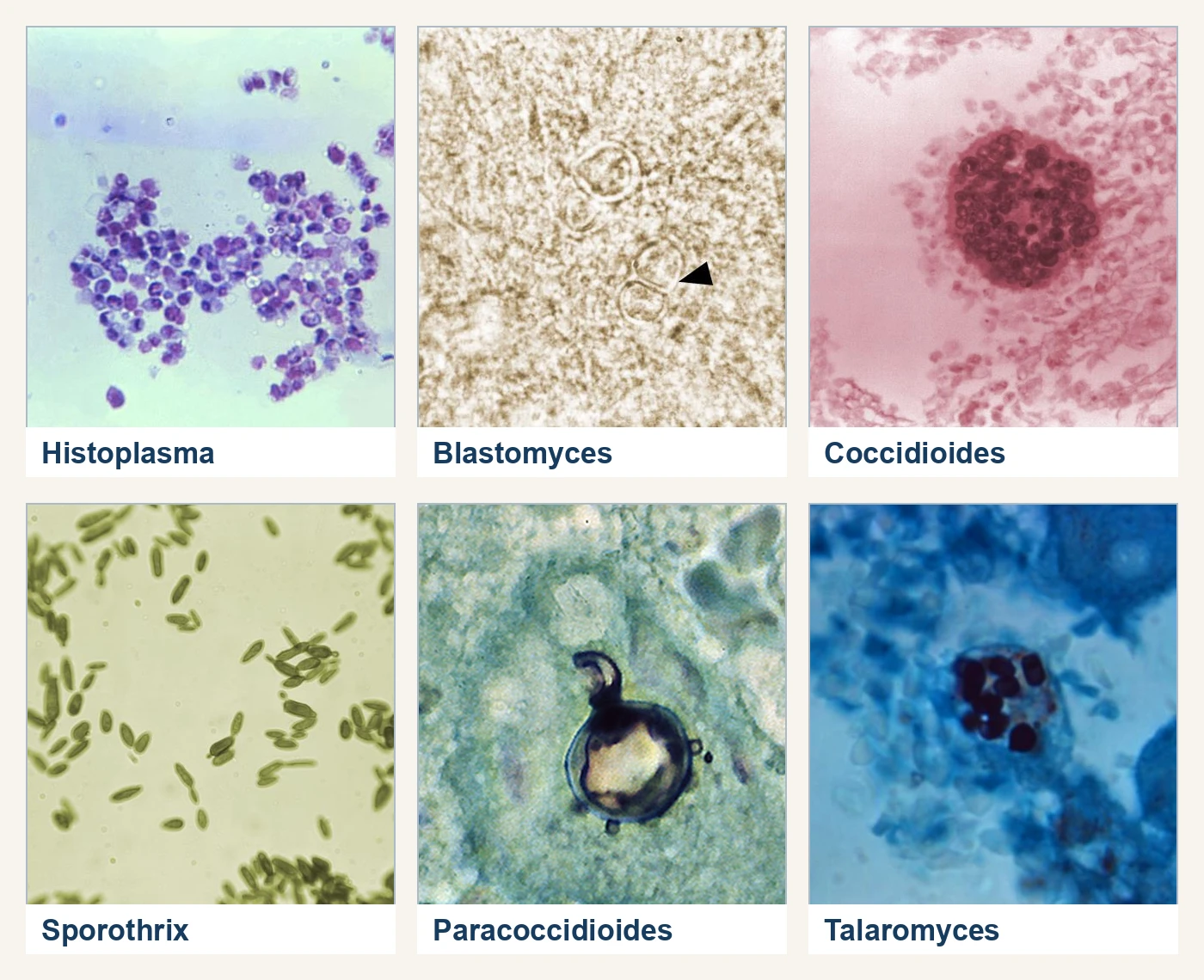
Handle suspected isolates with the containment and referral workflow described under handling fungal cultures safely: sealed plates, work only in a certified cabinet, no slide cultures, and identification by probe or referral with no morphologic manipulation. Treat an unidentified mold from a sterile site, a respiratory specimen, or a patient from an endemic area as a possible dimorphic pathogen until a probe result or a reference laboratory excludes it.
Practice
References
- Centers for Disease Control and Prevention. Identification of C. auris. Accessed August 31, 2026. https://www.cdc.gov/candida-auris/hcp/laboratories/identification-of-c-auris.html Back to text
- Centers for Disease Control and Prevention. Testing algorithm for histoplasmosis. Accessed August 31, 2026. https://www.cdc.gov/histoplasmosis/hcp/algorithm/index.html Back to text
- Centers for Disease Control and Prevention, National Institutes of Health. Biosafety in Microbiological and Biomedical Laboratories, 6th ed. CDC/NIH; revised June 2020. Accessed September 24, 2026. https://www.cdc.gov/labs/bmbl/index.html Back to text
- Federal Select Agent Program. HHS and USDA select agents and toxins. Accessed August 31, 2026. https://www.selectagents.gov/sat/list.htm Back to text
- Borman AM, Abdolrasouli A, Johnson EM. Name changes for fungi of medical importance, 2022-2024. J Clin Microbiol. 2025;63(11):e02041-24. doi:10.1128/JCM.02041-24 Back to text
- Clinical and Laboratory Standards Institute. Applying susceptibility interpretations to the Candida parapsilosis species complex. CLSI Insights. Accessed August 31, 2026. https://clsi.org/resources/insights-blog/applying-susceptibility-interpretations-to-the-candida-parapsilosis-species-complex/ Back to text
- Boulware DR, Rolfes MA, Rajasingham R, et al. Multisite validation of cryptococcal antigen lateral flow assay and quantification by laser thermal contrast. Emerg Infect Dis. 2014;20(1):45-53. doi:10.3201/eid2001.130906 Back to text
- Centers for Disease Control and Prevention. Clinical overview of cryptococcosis. Accessed August 31, 2026. https://www.cdc.gov/cryptococcosis/hcp/clinical-overview/index.html Back to text
- Ianiri G, LeibundGut-Landmann S, Dawson TL Jr. Malassezia: a commensal, pathogen, and mutualist of human and animal skin. Annu Rev Microbiol. 2022;76:757-782. doi:10.1146/annurev-micro-040820-010114 Back to text
- Pappas PG, Kauffman CA, Andes DR, et al. Clinical practice guideline for the management of candidiasis: 2016 update by the Infectious Diseases Society of America. Clin Infect Dis. 2016;62(4):e1-e50. doi:10.1093/cid/civ933 Back to text