In diagnostic parasitology, select the specimen and test for the suspected parasite and syndrome. A well-prepared specimen from the wrong site is unlikely to contain the parasite, and a negative result has little value when collection, transport, or test selection was poor. Suspected malaria and primary amebic meningoencephalitis are emergencies. The laboratory needs a round-the-clock plan that provides immediate testing or transfer without waiting for a routine parasitology shift.1
Specimen selection and preservation
Travel, residence, immune status, symptoms, exposure, treatment, and the suspected parasite should accompany the specimen. These details determine which concentration method, stain, antigen assay, molecular target, or reference laboratory is appropriate.
Scroll sideways to see all columns
| Clinical question | Preferred specimen | Handling that protects the result |
|---|---|---|
| Malaria | EDTA venous blood or fresh capillary blood; prepare at least 2 thick and 2 thin films | Make films as soon as possible, preferably within 1 hour. Collect during symptoms if practical, but never delay collection while waiting for a fever spike. |
| Babesiosis | EDTA whole blood with thin and thick films | Prepare films promptly. Retain whole blood refrigerated according to the molecular laboratory's instructions if polymerase chain reaction (PCR) testing may be needed. |
| African trypanosomiasis | Fresh blood, chancre fluid, lymph-node aspirate, or cerebrospinal fluid (CSF), selected by disease phase and exposure | Examine promptly. Concentration methods improve recovery when parasitemia is low. |
| Acute Chagas disease or reactivation | EDTA blood; tissue or other involved-site specimen when indicated | Use direct examination and a validated nucleic acid amplification test (NAAT). Chronic Chagas disease usually requires serum for serology. |
| Cutaneous or mucosal leishmaniasis | Scraping, brush sample, aspirate, touch preparation, or biopsy from active abnormal tissue | Before fixation, divide the specimen for direct smear, histopathology, culture, and molecular testing as required. Place culture material in the medium specified by the receiving laboratory. |
| Visceral leishmaniasis | Bone marrow aspirate is the usual first invasive specimen in North America; whole blood, buffy coat, lymph node, liver, or spleen may be appropriate | Splenic aspiration has high sensitivity and substantial bleeding risk. It is performed only in experienced centers. Prepare smears at the bedside when possible and reserve unfixed material for culture or NAAT. |
| Toxoplasmosis | Serum; amniotic fluid, CSF, blood, bronchoalveolar lavage, ocular fluid, placenta, or biopsy according to the syndrome | Follow the reference laboratory's temperature, volume, and transport requirements. Formalin-fixed tissue supports histology and immunohistochemistry but usually prevents culture. |
| Primary amebic meningoencephalitis | Fresh CSF and, when available, tissue | Keep the aliquot for immediate microscopy at room temperature. Send a separate unfixed aliquot for PCR according to the reference laboratory's instructions. |
| Amebic keratitis | Corneal scraping, biopsy, contact lens, lens case, and solution when requested | Inoculate culture media promptly and retain unfixed material for microscopy and PCR. |
| Granulomatous amebic encephalitis | Brain or skin biopsy; CSF is less sensitive | Submit both formalin-fixed tissue and unfixed tissue for histology, immunostaining, and PCR. |
Stool specimens
Liquid stool is more likely to contain fragile trophozoites. Formed stool is more likely to contain cysts. A fresh liquid specimen intended for motility examination should reach the laboratory within 30 minutes, and a soft specimen within 1 hour. When prompt microscopy is impossible, place portions into the preservatives required for concentration and permanent staining. Refrigeration is appropriate when the specific antigen or molecular assay accepts a refrigerated specimen, and formed stool may wait briefly, with overnight refrigeration if necessary, under the receiving laboratory's validated instructions. Refrigeration does not preserve fresh liquid or soft stool for the motile-trophozoite examination, which needs prompt processing or fixation.2
Collect stool into a clean, dry, leakproof container. Urine, water, soil, disinfectant, and absorbent material can destroy trophozoites or introduce artifacts. Barium and bismuth may interfere for 7 to 10 days, antimicrobial drugs for 2 to 3 weeks, and gallbladder contrast dyes for about 3 weeks. Antacids, kaolin, mineral oil, and nonabsorbable antidiarrheals can also obscure examination. Do not postpone an urgent diagnosis solely to meet those intervals. Document the interference and help select another method.
Scroll sideways to see all columns
| Preservative | Best use | Important limitation |
|---|---|---|
| 10% formalin | Concentration methods; recovery of many cysts, oocysts, eggs, and larvae; some special stains and immunoassays | Poor trophozoite detail; unsuitable for a routine trichrome smear; may inhibit some NAATs |
| Sodium acetate-acetic acid-formalin (SAF) | Concentration and permanent staining; compatible with some immunoassays | Morphology and assay compatibility vary with the staining system |
| Polyvinyl alcohol or Schaudinn-type fixative | Permanent stained smears with good intestinal protozoan detail | Poor choice for concentration, antigen testing, or NAAT; mercury-containing formulations require special handling |
| Legacy merthiolate-iodine-formalin (MIF) | Concentration and an iodine wet preparation | Rarely used in current practice; iodine can obscure or alter internal detail and limits compatibility with permanent stains and ancillary assays |
| Proprietary one-vial system | Concentration, permanent staining, and sometimes antigen testing or NAAT | Accept only the specimen types and methods named by the manufacturer and verified by the laboratory |
A conventional ova-and-parasite examination combines a concentration wet preparation with a permanent stained smear. It does not reliably cover every enteric parasite. Modified acid-fast or fluorescent stains may be needed for coccidia, modified trichrome or other dedicated methods for microsporidia, and antigen or molecular tests for selected targets. If microscopy is chosen and the first examination is negative, specimens collected on separate days, up to 3 in a series, improve recovery of intermittently shed organisms. Three specimens are not required in every case. A targeted antigen test or NAAT has its own collection instructions.1,2
Fresh and preserved stool remain biohazards. Fresh Ascaris and Trichuris eggs are usually unembryonated, and hookworm eggs contain a cleavage-stage embryo when promptly passed. Strongyloides stercoralis is usually recovered as a rhabditiform larva, although infective filariform larvae can develop during culture or prolonged delay. Fixation reduces risk but does not guarantee immediate inactivation of every cyst, oocyst, or egg.
Antigen, antibody, and molecular tests
Targeted tests detect fewer organisms than microscopy. A Giardia or Cryptosporidium antigen test does not detect helminths or other protozoa, and a multiplex gastrointestinal panel reports only the organisms included in that panel. Direct fluorescent-antibody assays can identify Giardia duodenalis cysts and Cryptosporidium oocysts. Their performance, accepted preservatives, centrifugation steps, and reporting rules are assay specific.
Serology is most useful when organisms are sparse, reside in deep tissue, or are no longer readily demonstrable. An antibody may reflect past exposure, cross-reaction, or persistent antibody after treated infection. Interpretation therefore depends on the organism and syndrome. Toxoplasmosis, babesiosis, Chagas disease, and visceral leishmaniasis each require a different serologic algorithm. Immunoglobulin class alone does not establish the stage of infection.
NAATs are available through commercial platforms, public health laboratories, and reference laboratories. They can improve sensitivity, distinguish morphologically overlapping species, and confirm an equivocal smear. Use the specimen type and intended use validated for that assay. A negative targeted NAAT excludes only the organisms and sequence variants the assay detects. Open amplification systems also require controls and physical or procedural separation that limits amplicon contamination.1
Blood-film preparation and examination
The thick and thin films serve different purposes. A thick film concentrates blood after erythrocytes lyse during staining, which makes it the more sensitive screening preparation. A thin film preserves erythrocyte shape, parasite architecture, and the relationship between the organism and its host cell. Species identification and percentage parasitemia depend mainly on the thin film.
- Collect capillary blood or venous blood into EDTA. For a fingerstick, wipe away the first drop and use freely flowing blood. Do not collect blood from an exposed venipuncture needle. EDTA is acceptable and practical, but storage progressively distorts parasites and erythrocytes.
- Prepare at least 2 thick and 2 thin films. A standardized thick film commonly uses about 6 µL spread into a circle 10 to 12 mm in diameter. Validate the volume, diameter, and drying time that give complete dehemoglobinization without washing the film from the slide.
- Air-dry films flat and protect them from insects, dust, heat, and formalin fumes. Fix only the thin film in absolute methanol. Methanol or heat fixation prevents the erythrocyte lysis needed in the thick film.
- Stain with a validated Romanowsky method. Giemsa in buffered water at approximately pH 7.2 remains the reference method for malaria because it preserves parasite chromatin, cytoplasm, pigment, and stippling. Wright or Wright-Giemsa can demonstrate parasites on a thin film when the procedure is validated. A methanol-containing stain must not fix the thick film before dehemoglobinization.
- Scan both preparations at 10× or 20× for distribution, stain quality, and large blood parasites. Examine the thick film under 100× oil in an evenly stained area containing roughly 10 to 20 leukocytes per field. Examine the thin film in the monolayer where erythrocytes touch with little overlap. Cells at the extreme feathered edge are distorted.
- For suspected malaria, examine at least 100 thick-film oil-immersion fields before reporting a negative conventional examination. A laboratory may require 200 to 300 fields or the whole usable film when parasite density is expected to be low. Follow the validated procedure and document the extent of review.3
Stain precipitate is irregular and usually lies above or between cells. A platelet over an erythrocyte may resemble a chromatin dot but lacks a surrounding parasite cytoplasm. Howell-Jolly bodies are round, dense nuclear remnants without blue parasite cytoplasm. A true malaria or Babesia call requires a reproducible population of organisms with internally consistent structure.
A calibrated ocular micrometer is useful whenever size helps narrow an identification, especially for stool protozoa, oocysts, eggs, larvae, and tissue forms. Calibrate each objective used for measurement. Size supports identification together with nuclear structure, cytoplasmic features, host-cell changes, specimen source, and clinical context.
Peripheral blood concentration methods have narrower roles. A buffy-coat preparation can increase recovery of trypanosomes and microfilariae. The Knott method and membrane filtration concentrate selected microfilariae.
Malaria
Female Anopheles mosquitoes transmit human malaria naturally. Malaria can also be acquired congenitally or through transfusion, transplantation, shared needles, or laboratory exposure. Six taxa are established causes of human malaria: Plasmodium falciparum, P. vivax, P. malariae, P. knowlesi, P. ovale curtisi, and P. ovale wallikeri. The two P. ovale species are reported together as the P. ovale complex when they have not been distinguished molecularly.4,5
The mosquito injects sporozoites, the stage infective to humans. Hepatic schizogony (asexual multiplication inside liver cells) releases merozoites, which invade erythrocytes and develop through ring, growing trophozoite, and schizont stages. Some become gametocytes, the stage infective to the mosquito. The mosquito is the definitive host because sexual reproduction occurs there. Humans are intermediate hosts. P. vivax and the P. ovale complex form dormant hepatic hypnozoites that can cause relapse. Every malaria species can persist or recur from blood-stage infection, termed recrudescence.
The approximate erythrocytic cycles are 24 hours for P. knowlesi, 48 hours for P. falciparum, P. vivax, and the P. ovale complex, and 72 hours for P. malariae. Clinical fever patterns are often irregular, especially early in disease, in mixed infection, or after antimalarial exposure. Never make the laboratory species call from periodicity.
Emergency testing sequence
A patient with fever or compatible illness and possible exposure in a malaria-endemic area requires an immediate thick and thin film. Report the presence of parasites, the best-supported species identification, and parasitemia as soon as each is available. If the first films are negative and malaria remains possible, repeat films every 12 to 24 hours for a total of 3 sets. A rapid, laboratory-validated molecular test may run concurrently and can shorten that sequence when its negative result has been validated to exclude malaria in the tested population. Reference PCR is especially useful for low-density infection, mixed infection, and species overlap, but a send-out result must not delay initial microscopy.4,6
Species morphology on thin films
Scroll sideways to see all columns
| Feature | P. falciparum | P. vivax | P. ovale complex | P. malariae | P. knowlesi |
|---|---|---|---|---|---|
| Infected erythrocyte | Usually normal size; multiple infection is common | Normal to about 1.25× in early rings, then often 1.5-2× | Normal to about 1.25×; may become oval with fimbriated edges | Normal to smaller, sometimes about 0.75× | Usually normal to smaller, sometimes about 0.75× |
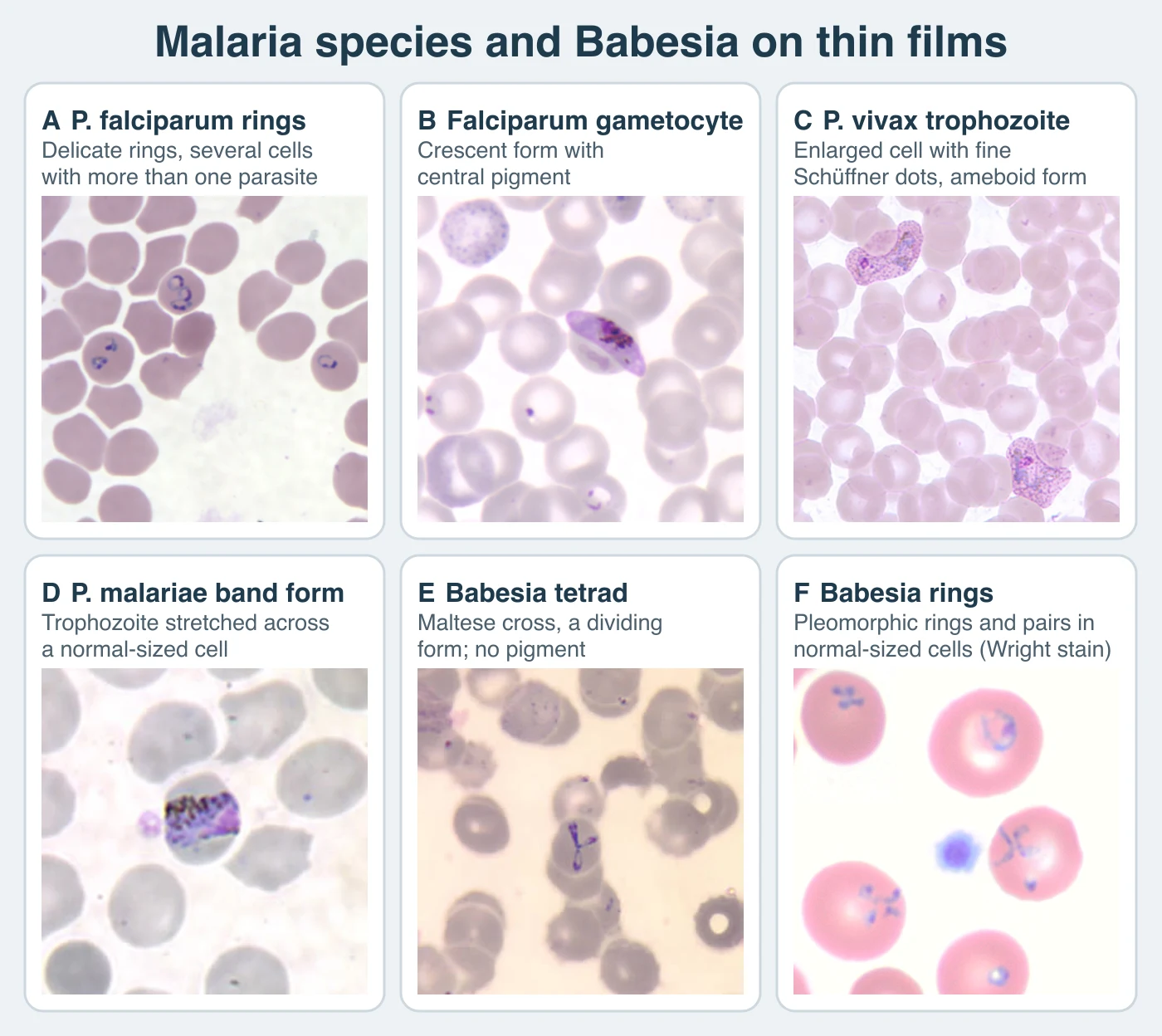
| Young trophozoite | Very delicate ring, about one-fifth of the erythrocyte diameter; double chromatin dots and appliqué forms may occur | Larger ring with one chromatin dot; later cytoplasm becomes irregular and ameboid | Compact ring; later trophozoite remains more compact than P. vivax | Compact ring; later trophozoite may stretch across the cell as a band | Early rings may resemble P. falciparum, including double dots, appliqué position, and multiple infection; later bands resemble P. malariae |
| Stippling or clefts | Coarse, irregular Maurer clefts may be visible | Fine Schüffner dots become prominent as the cell enlarges | James dots, also called Schüffner-like stippling, are often conspicuous | Fine Ziemann dots are uncommon and easy to miss | Fine Sinton and Mulligan stippling may occur but is not a reliable stand-alone discriminator |
| Schizont | Usually sequestered; when present, about 8-24 merozoites around dark pigment | About 12-24 merozoites, commonly 16 | About 6-14 merozoites, commonly 8 | About 6-12 merozoites, commonly 8; a rosette around central pigment is characteristic | Up to about 16 merozoites; mature forms can closely resemble P. malariae |
| Gametocyte | Mature form is crescent or banana shaped; immature forms sequester | Round to oval, filling an enlarged erythrocyte | Round to oval in a stippled erythrocyte | Round to oval in a normal-sized erythrocyte | Round to oval and morphologically overlaps other non-falciparum species |
| Circulating pattern | Rings and mature gametocytes predominate; mature trophozoites and schizonts are uncommon but can appear, especially with high parasite burden or delayed processing | All blood stages may circulate | All blood stages may circulate | All blood stages may circulate | All blood stages may circulate |
Base the species identification on all the forms present on the film. Delayed film preparation can compact or distort parasites. Early P. knowlesi stages can resemble P. falciparum. Later stages can resemble P. malariae. Report a provisional identification and obtain species-level PCR when exposure makes P. knowlesi possible. Molecular testing also resolves morphologically indistinguishable P. ovale species, low-density mixed infections, and conflicting morphology. A mature P. falciparum trophozoite or schizont in peripheral blood is concerning, but that finding alone does not define severe malaria.5
Parasitemia
Report quantitative parasitemia for every smear-positive malaria case. Percentage parasitemia from the thin film is:
Percent infected erythrocytes = infected erythrocytes ÷ erythrocytes counted × 100
Count 500 to 2,000 erythrocytes in representative monolayer fields, following your laboratory's validated procedure. Count an erythrocyte containing several asexual parasites once. Exclude gametocytes from the asexual parasitemia. For example, 96 infected cells among 2,400 erythrocytes gives 4.0% parasitemia.
Estimate asexual parasite density from the thick film when percentage parasitemia cannot be determined reliably:
Parasites/µL = asexual parasites counted ÷ leukocytes counted × patient leukocyte count/µL
Use the patient's measured leukocyte count whenever available. A common endpoint is 500 parasites or 1,000 leukocytes, whichever comes first. If the procedure substitutes 8,000 leukocytes/µL, state that assumption in the report. A count of 125 parasites per 500 leukocytes with a measured leukocyte count of 6,000/µL gives 1,500 parasites/µL. Repeat quantitation during treatment at the interval required by the clinical protocol, using the same validated method when possible.3,4
Rapid antigen and molecular testing
As of August 2026, BinaxNOW Malaria is the only malaria rapid diagnostic test cleared by the Food and Drug Administration (FDA) in the United States. It detects P. falciparum histidine-rich protein 2 (HRP2) and a pan-Plasmodium aldolase. It does not provide a reliable species identification for every non-falciparum infection, quantify parasitemia, or monitor clearance. Sensitivity falls at low parasite density and with some non-falciparum infections. pfhrp2 or pfhrp3 deletion can produce an HRP2-negative P. falciparum result. HRP2 can remain detectable for weeks after parasites clear. Every positive or negative rapid antigen result requires correlation with immediate thick and thin films.6,7
PCR and other NAATs are more sensitive than conventional microscopy in many settings and are valuable for species confirmation. Clinical diagnostic assays and blood-donor screening assays have different intended uses. Do not treat a donor-screening nucleic acid test (NAT) as a patient diagnostic test unless that use has been separately validated.
Babesiosis
Babesia species are intraerythrocytic apicomplexans transmitted by ticks. Transfusion is a well-established route, and congenital transmission occurs rarely. In the northeastern and upper midwestern United States, B. microti cycles mainly between Ixodes scapularis and the white-footed mouse, Peromyscus leucopus. Rare western US infections are caused by B. duncani. Current evidence supports Dermacentor albipictus as its vector and mule deer as an important reservoir. Do not assign Ixodes pacificus as the vector for B. duncani. European human babesiosis includes B. divergens and B. venatorum, commonly associated with Ixodes ricinus.8,9
The tick injects sporozoites, which enter erythrocytes directly without a hepatic stage. Asexual trophozoites and merozoites are the diagnostic forms. Some parasites become gametes that can continue the sexual cycle after ingestion by the tick, the definitive host, but those gametes cannot be distinguished by routine light microscopy. Humans usually do not maintain the natural enzootic cycle.
Blood-film findings
Babesia trophozoites range from tiny delicate rings to pleomorphic, vacuolated, ameboid, or pyriform forms. Infected erythrocytes remain normal in size. Multiple infection, paired pear-shaped forms, and occasional extracellular parasites support babesiosis. Pigment is absent. A four-member tetrad, the Maltese cross, is diagnostic when present but is uncommon. Its absence does not exclude babesiosis. Species cannot be assigned by morphology alone.8
Scroll sideways to see all columns
| Feature | Babesia spp. | P. falciparum |
|---|---|---|
| Erythrocyte size | Normal | Usually normal |
| Ring population | Markedly pleomorphic; pyriform pairs and extracellular forms may occur | Delicate rings are more uniform; appliqué forms and double chromatin dots may occur |
| Pigment | Absent | Hemozoin appears in mature stages and gametocytes |
| Tetrad | Maltese cross is diagnostic but rare | Absent |
| Recognizable gametocyte | No Plasmodium-type gametocyte | Crescent mature gametocyte supports P. falciparum |
| Nonerythrocytic stage in humans | None | Hepatic stage precedes erythrocytic infection |
Examine both thick and thin films, recognizing that tiny Babesia forms may be difficult to see in a dehemoglobinized thick film. Review a broad area of the thin film, often 200 to 300 oil-immersion fields in a low-density infection under a validated procedure. If films are negative and suspicion persists, use PCR. A species-specific B. microti PCR can miss another Babesia species, so exposure in the western United States, Europe, or another region may call for a pan-Babesia assay or additional species targets. If travel and morphology leave malaria in the differential, pursue molecular testing for both groups without waiting to find a Maltese cross.
Calculate percent infected erythrocytes with the same thin-film equation used for malaria. Quantitation establishes baseline burden and helps follow response in selected patients. A single positive antibody result does not prove active babesiosis because antibody can persist. A 4-fold rise in B. microti IgG in paired sera supports recent infection. Smear or PCR establishes current parasitemia. Blood-donor antibody and NAT screening protects the blood supply. Those assays are not interchangeable with clinical diagnostic tests.10
Hemoflagellates
Trypanosoma and Leishmania are kinetoplastids. Their kinetoplast is a compact mass of mitochondrial DNA that stains as a dark red-purple dot or short rod near the nucleus on a Romanowsky-stained preparation.
Scroll sideways to see all columns
| Form | Diagnostic morphology | Usual location and significance |
|---|---|---|
| Amastigote | Round to oval; nucleus and separate kinetoplast; no external flagellum | Intracellular form of Leishmania and T. cruzi in mammalian tissue |
| Promastigote | Elongated; kinetoplast anterior to the nucleus; one free anterior flagellum; no undulating membrane | Leishmania in the sand fly and culture; the metacyclic promastigote infects the mammal |
| Epimastigote | Elongated; kinetoplast just anterior to the nucleus; short undulating membrane and free flagellum | Replicative form of trypanosomes in the vector or in culture, so it is rarely encountered in human diagnostic specimens |
| Trypomastigote | Elongated; central nucleus, posterior kinetoplast, undulating membrane, and free flagellum | Diagnostic bloodstream form of T. brucei and T. cruzi; the metacyclic form infects the mammal |
African trypanosomiasis
A tsetse fly in the genus Glossina injects metacyclic trypomastigotes. Bloodstream trypomastigotes multiply by longitudinal binary fission in blood, lymph, and later CSF. Trypanosoma brucei gambiense occurs in West and Central Africa, usually causes a slowly progressive illness, and has humans as its principal reservoir, although animals can be infected. T. b. rhodesiense occurs in eastern and southern Africa, usually progresses more rapidly, and is a zoonosis maintained in cattle and wildlife.11,12
The two subspecies are morphologically indistinguishable. Trypomastigotes are approximately 14 to 33 µm long, with a small posterior kinetoplast, central nucleus, undulating membrane, and free flagellum. Slender and stumpy forms may occur, and dividing forms can be seen. T. b. rhodesiense often produces a higher parasitemia, so fresh blood, thick and thin films, or a buffy-coat concentration may show organisms readily. Gambiense disease has a lower parasitemia and often requires repeated examination and concentration.
Confirmation requires demonstration of the parasite in chancre fluid, lymph-node aspirate, blood, bone marrow in selected cases, or CSF. Antibody rapid tests and the card agglutination test for trypanosomiasis screen for T. b. gambiense in appropriate populations. They do not confirm infection and do not diagnose T. b. rhodesiense. Where treatment selection still requires CSF staging, trypanosomes in CSF or more than 5 leukocytes/µL supports second-stage disease. A patient with no CSF trypanosomes and 5 or fewer leukocytes/µL meets the conventional first-stage laboratory definition. Molecular assays are specialized adjuncts and are not used alone for routine confirmation.
Chagas disease
Triatomine insects transmit Trypanosoma cruzi. Infective metacyclic trypomastigotes are passed in bug feces and enter through the bite site, conjunctiva, or another mucosal surface. Oral, congenital, transfusion, transplantation, and laboratory transmission also occur. In mammalian cells the parasite becomes an amastigote, divides, and differentiates into a bloodstream trypomastigote. Bloodstream forms circulate without dividing.
Acute infection, congenital infection, reactivation during immunosuppression, and some orally acquired outbreaks can produce detectable parasitemia. A T. cruzi trypomastigote is about 12 to 30 µm long, often curves into a C or U shape, and has a large terminal or subterminal kinetoplast, central nucleus, undulating membrane, and free flagellum. Shape alone is not specific. Tissue amastigotes occur in clusters and cannot be reliably separated from Leishmania amastigotes without epidemiologic and ancillary evidence.
Use fresh blood concentration, stained blood films, and a validated PCR for acute, congenital, or reactivated disease. Chronic infection usually has parasitemia below the sensitivity of microscopy and intermittently below the sensitivity of PCR. Diagnosis therefore requires positive results from 2 serologic assays that use different antigens or formats. Use a third assay to resolve discordant results. A negative blood PCR does not exclude chronic Chagas disease. Chronic cardiomyopathy and gastrointestinal disease reflect persistent infection with inflammatory injury to myocardium, smooth muscle, and the autonomic nervous system. Current evidence favors this mechanism over a purely autoimmune loss of parasympathetic cells.13
Leishmaniasis
Female phlebotomine sand flies transmit Leishmania: Phlebotomus in the Old World and several New World genera historically grouped as Lutzomyia. Reservoirs vary by species and include humans, dogs, and wild mammals. The metacyclic promastigote infects the mammal. Macrophages ingest it, and intracellular amastigotes multiply in the phagolysosomal compartment. The sand fly ingests amastigotes during a blood meal, and promastigotes develop in the fly.
Only amastigotes occur in human tissue. They are usually 1 to 5 µm long by 1 to 2 µm wide and contain a nucleus and a smaller rod-shaped kinetoplast. Organisms may be extracellular when cells rupture. Species are morphologically indistinguishable. T. cruzi amastigotes have the same basic structure. Histoplasma yeasts lack a kinetoplast and may show narrow-based budding. Gomori methenamine silver (GMS) stain highlights fungal cell walls. Fungal stains do not reliably stain Leishmania.
Cutaneous and mucosal disease require sampling from active abnormal tissue. Prepare direct smears or touch preparations, histopathology, culture, and molecular testing as the specimen allows. Visceral disease is associated chiefly with the L. donovani complex, including L. donovani and L. infantum. The latter was historically reported in the Americas as L. chagasi. Bone marrow is commonly sampled first in North America. Splenic aspirate has the highest sensitivity but also a serious hemorrhage risk. Blood or buffy coat may be useful, especially in immunocompromised patients.
Microscopy or histopathology can establish infection when characteristic amastigotes are demonstrated. Culture in Novy-MacNeal-Nicolle medium or a suitable tissue-culture medium yields promastigotes and may require up to 4 weeks of incubation. PCR improves sensitivity and enables species identification, which can affect prognosis and management. Serology supports visceral leishmaniasis in an immunocompetent patient but performs poorly for cutaneous disease and may be falsely negative in patients with HIV. A positive antibody result does not prove active visceral infection in an endemic population.14
Toxoplasma gondii
Felids are the definitive hosts of Toxoplasma gondii. They shed unsporulated oocysts, which usually become infective after 1 to 5 days in the environment. A mature oocyst is about 10 to 12 µm and contains 2 sporocysts, each with 4 sporozoites. Humans acquire infection by ingesting sporulated oocysts or tissue cysts in undercooked meat. Congenital, transplantation-associated, transfusion-associated, and laboratory transmission also occur.
Tachyzoites are crescent shaped, approximately 4 to 8 µm by 2 to 3 µm, and multiply during active dissemination. Tissue cysts are commonly 5 to 50 µm or larger and contain numerous slender bradyzoites. A bradyzoite is the slow-growing organism inside the tissue cyst. Each cyst contains many bradyzoites. Tissue cysts persist in neural and muscle tissue and can reactivate during immunosuppression.15
Diagnostic patterns
- Immunocompetent patient: Begin with serum IgG and IgM when recent systemic infection is suspected. IgG documents infection at some time. IgM may persist for 18 months or longer and can be falsely positive, so a result used to date infection requires confirmation by a toxoplasmosis reference laboratory. High IgG avidity early in pregnancy can exclude infection acquired within the assay's stated recent interval. Low avidity does not prove recent infection.
- Pregnancy and fetus: Interpret maternal IgG, IgM, confirmatory serology, avidity, and serial specimens together. PCR of amniotic fluid collected at the appropriate gestational interval detects many fetal infections. Maternal blood PCR is not the routine test for fetal infection.
- Newborn: Maternal IgG crosses the placenta, so neonatal IgG alone cannot establish congenital infection. Reference-laboratory IgM and IgA, comparison of maternal and infant antibody profiles, PCR or direct detection when available, and serial infant IgG are used together. Persistence or rise of infant IgG beyond the expected loss of maternal antibody supports congenital infection.
- Immunocompromised patient: Toxoplasmic encephalitis usually represents reactivation. Serum IgG is commonly positive and IgM commonly absent. CSF PCR is highly specific but has limited sensitivity, especially after treatment begins. A negative result does not exclude encephalitis. Demonstration of tachyzoites or tissue cysts by histology, immunohistochemistry, or PCR in CSF, blood, bronchoalveolar lavage, ocular fluid, or biopsy can establish disease in the appropriate syndrome.
Earlier maternal infection is less likely to transmit across the placenta but more likely to cause severe fetal injury when transmission occurs. Transmission becomes more frequent later in gestation. Many congenitally infected infants appear well at birth. Report wording should not imply that an asymptomatic newborn is uninfected or that a single antibody class shows whether or when infection occurred.
Free-living amebae
Scroll sideways to see all columns
| Organism | Disease and exposure | Morphology in clinical material | Principal laboratory approach |
|---|---|---|---|
| Naegleria fowleri | Primary amebic meningoencephalitis after warm fresh water or inadequately disinfected water enters the nose; unsafe nasal irrigation is another route | Trophozoite 10-35 µm, often vacuolated, with one nucleus and a large central karyosome without peripheral chromatin. A temporary flagellate form may rarely be seen in CSF. Cysts do not occur in human tissue. | Immediate wet preparation of fresh room-temperature CSF; stained cytospin or smear; PCR and immunohistochemistry or immunofluorescence through a reference laboratory |
| Acanthamoeba spp. | Keratitis, often associated with contact lenses or corneal trauma; granulomatous amebic encephalitis and disseminated disease, usually with immune compromise | Trophozoite 15-45 µm with spine-like acanthopodia; cyst 10-25 µm with a wrinkled outer wall and polygonal, stellate, or round inner wall. Both forms may occur in tissue. | Corneal scraping microscopy, culture, and PCR for keratitis; tissue histology, PCR, and immunostaining for encephalitis. In vivo confocal microscopy is an adjunct for keratitis. |
| Balamuthia mandrillaris | Granulomatous amebic encephalitis and skin disease in immunocompetent or immunocompromised people; entry through skin or respiratory tract with hematogenous spread; donor-derived transmission can occur | Trophozoite about 15-60 µm with broad pseudopodia; cyst commonly 10-25 µm. The cyst appears double walled by light microscopy and has a three-layer wall ultrastructurally. Both forms may occur in tissue. | Tissue histology with species-specific PCR, immunohistochemistry, or immunofluorescence. Routine light microscopy cannot reliably separate it from Acanthamoeba. |
Suspected primary amebic meningoencephalitis requires immediate action. Examine fresh CSF at room temperature for motile trophozoites and notify the clinical team of a presumptive finding at once. A stained preparation may resemble neutrophils, so look for the large karyosome, vacuolated cytoplasm, and pseudopod formation. Gram-stain heat fixation can damage organisms. Preserve a separate unfixed aliquot for PCR and contact public health authorities or the Centers for Disease Control and Prevention (CDC) for emergency diagnostic assistance. A negative wet preparation does not exclude infection.16
An annular or ring corneal infiltrate supports Acanthamoeba keratitis but often appears late and is not required. Corneal scrapings can be examined with calcofluor white, Giemsa, trichrome, or another validated stain and cultured on nonnutrient agar overlaid with bacteria. Balamuthia does not grow on this routine amebic culture system. Specialized mammalian-cell culture is confined to experienced laboratories. Brain or skin tissue generally has a higher yield than CSF for Acanthamoeba or Balamuthia encephalitis. Handle cultures and unfixed specimens with biosafety level 2 (BSL-2) practices and organism-specific risk controls.17
Quality and reporting
Run positive, negative, and internal controls with special stains, antigen assays, and NAATs at the frequency required by each method. New reagent lots need performance checks. Reference slides and image atlases support morphology review. Unknowns and competency specimens assess whether the examiner can recognize low-density and mixed populations. Ocular micrometers require objective-specific calibration.
Name the organism only to the level the evidence supports. When the evidence does not support a species, report a provisional Plasmodium or kinetoplastid identification and state the differential, urgency, and confirmation plan. Preserve representative positive films, document the fields or cells counted, and arrange prompt expert or public health review when the morphology is unusual, the parasite is rare, or the result conflicts with exposure history. At the bench, treat every malaria request as a stat test, from the first film to the parasitemia.
Practice
References
- Miller JM, Binnicker MJ, Campbell S, et al. A guide to utilization of the microbiology laboratory for diagnosis of infectious diseases: 2024 update by the Infectious Diseases Society of America and the American Society for Microbiology. Clin Infect Dis. 2024;ciae104. doi:10.1093/cid/ciae104 Back to text
- Centers for Disease Control and Prevention. DPDx: stool specimens, specimen collection. Accessed August 31, 2026. https://www.cdc.gov/dpdx/diagnosticprocedures/stool/specimencoll.html Back to text
- Centers for Disease Control and Prevention. DPDx: blood specimens, specimen processing, staining, and microscopic examination. Accessed August 31, 2026. https://www.cdc.gov/dpdx/diagnosticprocedures/blood/index.html Back to text
- Centers for Disease Control and Prevention. Evaluation and diagnosis of malaria. Accessed August 31, 2026. https://www.cdc.gov/malaria/hcp/clinical-guidance/evaluation-diagnosis.html Back to text
- Centers for Disease Control and Prevention. DPDx: malaria. Accessed August 31, 2026. https://www.cdc.gov/dpdx/malaria/ Back to text
- Centers for Disease Control and Prevention. Malaria diagnostic tests. Accessed August 31, 2026. https://www.cdc.gov/malaria/hcp/diagnosis-testing/malaria-diagnostic-tests.html Back to text
- US Food and Drug Administration. BinaxNOW Malaria: de novo classification request DEN070006. Accessed August 31, 2026. https://www.accessdata.fda.gov/scripts/cdrh/cfdocs/cfpmn/denovo.cfm?id=DEN070006 Back to text
- Centers for Disease Control and Prevention. DPDx: babesiosis. Accessed August 31, 2026. https://www.cdc.gov/dpdx/babesiosis/index.html Back to text
- Swei A, O'Connor KE, Couper LI, et al. Evidence for transmission of the zoonotic apicomplexan parasite Babesia duncani by the tick Dermacentor albipictus. Int J Parasitol. 2019;49(2):95-103. doi:10.1016/j.ijpara.2018.07.002 Back to text
- Krause PJ, Auwaerter PG, Bannuru RR, et al. Clinical practice guidelines by the Infectious Diseases Society of America: 2020 guideline on diagnosis and management of babesiosis. Clin Infect Dis. 2021;72(2):e49-e64. doi:10.1093/cid/ciaa1216 Back to text
- Centers for Disease Control and Prevention. DPDx: African trypanosomiasis. Accessed August 31, 2026. https://www.cdc.gov/dpdx/trypanosomiasisafrican/index.html Back to text
- World Health Organization. Trypanosomiasis, human African (sleeping sickness). Accessed August 31, 2026. https://www.who.int/news-room/fact-sheets/detail/trypanosomiasis-human-african-%28sleeping-sickness%29 Back to text
- Centers for Disease Control and Prevention. Clinical testing and diagnosis for Chagas disease. Accessed August 31, 2026. https://www.cdc.gov/chagas/hcp/diagnosis-testing/index.html Back to text
- Centers for Disease Control and Prevention. DPDx: leishmaniasis. Accessed August 31, 2026. https://www.cdc.gov/dpdx/leishmaniasis/index.html Back to text
- Centers for Disease Control and Prevention. DPDx: toxoplasmosis. Accessed August 31, 2026. https://www.cdc.gov/dpdx/toxoplasmosis/index.html Back to text
- Centers for Disease Control and Prevention. Clinical and laboratory diagnosis for Naegleria fowleri infection. Accessed August 31, 2026. https://www.cdc.gov/naegleria/hcp/diagnosis-testing/index.html Back to text
- Centers for Disease Control and Prevention. DPDx: free-living amebic infections. Accessed August 31, 2026. https://www.cdc.gov/dpdx/freelivingamebic/index.html Back to text