Most respiratory specimens pass through heavily colonized airways before reaching the laboratory, so recovering an organism does not establish that it is a pathogen. Workup and reporting depend on whether the specimen represents the stated site and whether recovered organisms exceed expected colonization. Pathogens that do not grow on routine media require a specific request or molecular assay. General transport rules, Gram-stain mechanics, media, incubation, and the staged reporting cycle are covered in Specimen Collection, Transport, and Primary Processing in Microbiology.
Selecting lower respiratory specimens for culture
Sputum culture has limited value in mild community-acquired pneumonia, so it is ordered for selected patients. In hospitalized adults, a pretreatment lower respiratory Gram stain and culture is recommended for severe pneumonia, for patients being treated empirically for methicillin-resistant Staphylococcus aureus (MRSA) or Pseudomonas aeruginosa, for patients with prior respiratory isolation of either organism, and for patients hospitalized and given parenteral antimicrobials within the previous 90 days. When the patient is intubated, an endotracheal aspirate collected promptly after intubation gives better yield than expectorated sputum. Clinicians use negative results from these cultures when they review broad empiric coverage.1
Lower respiratory specimens include expectorated or induced sputum, tracheal aspirate, bronchoalveolar lavage (BAL) fluid, protected-brush specimens, and bronchial washings.2,3
Expectorated sputum is best collected early in the morning before eating, after the patient rinses the mouth with water and produces a deep-cough specimen of 5 to 10 mL. A nonproductive cough can be induced with aerosolized saline. An induced specimen is thin and watery and can be mistaken for saliva on appearance alone, so the requisition identifies it as induced and the laboratory screens it accordingly.3
Sputum collected for mycobacterial culture follows different rules. At least three specimens are collected, 8 to 24 hours apart, with at least one taken early in the morning.4 The patient must not rinse the mouth with tap water first, because a water rinse can introduce environmental mycobacteria such as Mycobacterium gordonae or the M. avium-intracellulare complex and seed the specimen with organisms that can be mistaken for a true pathogen.3
Screening sputum for adequacy
Sputum passes through the oropharynx, so a screening Gram stain assesses whether it contains lower respiratory secretions or mostly saliva. Apply your laboratory's adopted screening criteria. The routine bacterial culture policy used here rejects specimens with more than 25 squamous epithelial cells per low-power field because they indicate salivary contamination. A specimen with 25 or fewer passes this cellular screen if the other specimen checks are acceptable. This policy does not require a minimum neutrophil count. Record a rejection and its reason, and request a fresh specimen.17
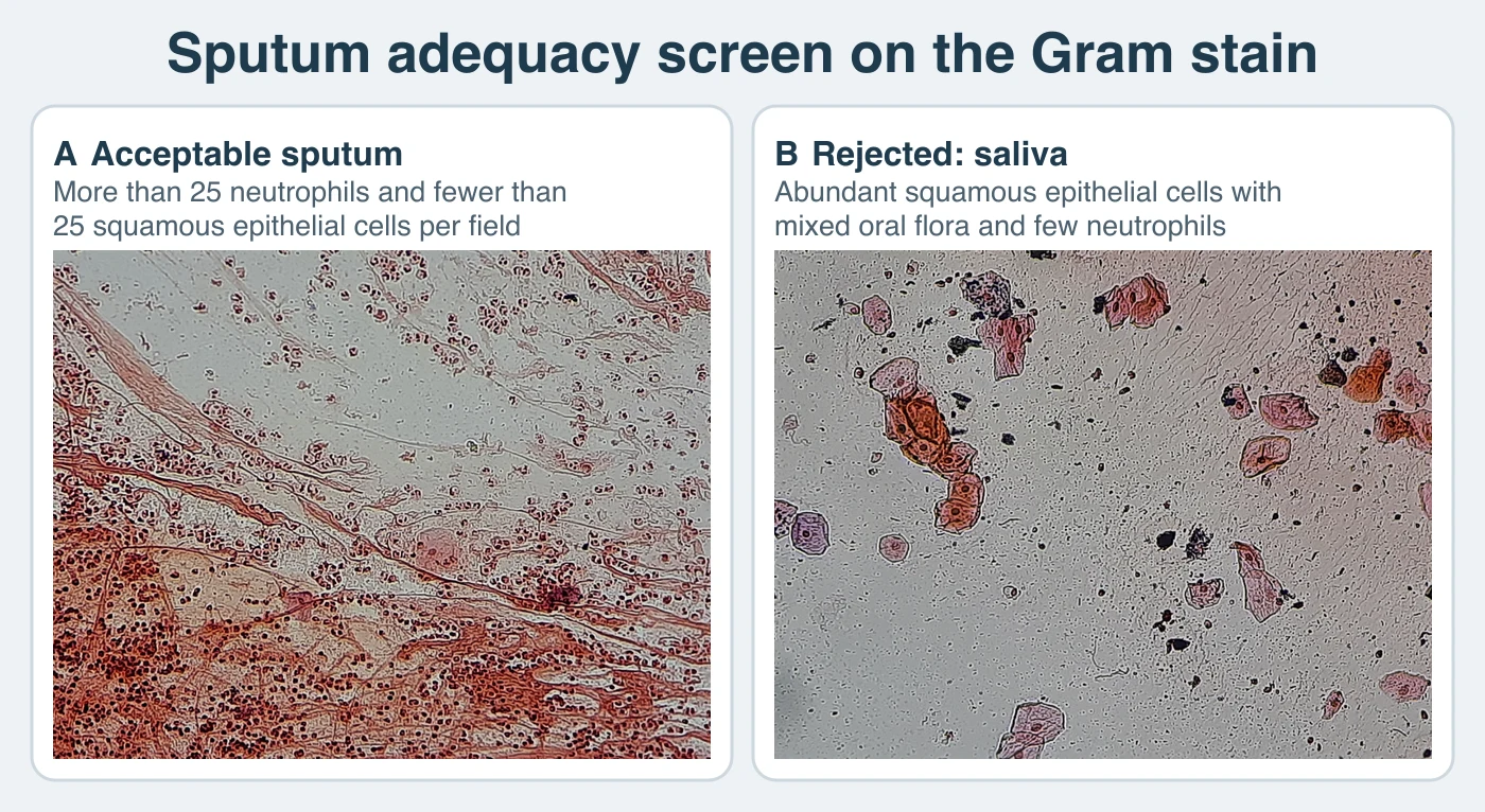
Neutrophil numbers alone never determine acceptability. A neutropenic patient can have bacterial pneumonia with few neutrophils in the specimen.3
The screen applies only to routine bacterial culture. Mycobacterial, fungal, and Pneumocystis requests are processed regardless of the squamous cell count. For suspected legionellosis, do not reject a lower respiratory specimen because it appears contaminated or contains few leukocytes, since Legionella recovery does not depend on those features.5,3
Indigenous flora of the mouth and upper airway
Recognizing indigenous oral and upper-airway flora prevents colonizers from being worked up as pathogens. These organisms account for most growth on throat cultures.
Scroll sideways to see all columns
| Resident organism | Gram-stain appearance | Culture note |
|---|---|---|
| Viridans streptococci | Gram-positive cocci in chains | Small alpha-hemolytic colonies |
| Neisseria species other than N. gonorrhoeae and N. meningitidis | Gram-negative diplococci | Common nasopharyngeal residents |
| Moraxella catarrhalis | Gram-negative diplococci | Nasopharyngeal carriage in healthy children and adults; also a pathogen |
| Coagulase-negative staphylococci | Gram-positive cocci in clusters | Ubiquitous skin and mucosal flora |
| Corynebacterium species (diphtheroids) | Gram-positive rods in palisades and V forms | Nonhemolytic, dry colonies |
| Haemophilus species | Small gram-negative coccobacilli | Resident and pathogen, so significance depends on the clinical picture |
| Candida species | Budding yeast, sometimes with pseudohyphae | Part of resident oral flora |
| Prevotella species | Small gram-negative rods | Oral anaerobe |
| Fusobacterium species | Gram-negative rods with thin, pointed ends | Oral anaerobe |
| Veillonella species | Small gram-negative cocci | Oral anaerobe |
| Actinomyces species | Branching, filamentous gram-positive rods | Oral anaerobe |
Mixed flora of this kind is expected in specimens that pass through the mouth and does not alone make the specimen unacceptable. Squamous epithelial cells are the best marker of oral contamination. Numerous squamous cells with mixed flora and few or no neutrophils indicate saliva.3
Bronchoscopy specimens
BAL fluid and protected-brush specimens are used for acute bacterial pneumonia, for ventilator-associated pneumonia in patients not yet treated, for opportunistic pathogens in immunocompromised patients, and for mycobacterial infection when a patient cannot produce sputum.3
A protected-brush specimen is collected through a double-cannula catheter holding only 0.001 to 0.01 mL of secretions. A displaceable plug seals the outer cannula until it reaches the sampling site. The brush and inner cannula retract into it before withdrawal to prevent upper-airway flora from contaminating the brush during passage. Place the brush in 1 mL of sterile saline or broth for transport.3
BAL is performed by wedging the bronchoscope tip into an airway and instilling sterile saline in three or four aliquots, commonly more than 140 mL in total, which samples an estimated one million alveoli. Between 10 and 100 mL usually returns. The fluid should reach the laboratory within 30 minutes, because carryover from the procedure inhibits some bacteria. When a delay is unavoidable, refrigeration limits that loss only partly.3
Vortex BAL fluid for culture inoculation and cytocentrifuge preparations. Keep small tissue fragments moist in sterile saline and process them separately. Treat viscous fluid suspected of harboring fungi, including Pneumocystis jirovecii, with a mucolytic agent and centrifuge it before staining. A Gram-stained cytospin showing one or more bacteria per oil-immersion field and no squamous epithelial cells strongly suggests acute bacterial pneumonia. A Papanicolaou-stained cytospin can show the cytopathic inclusions of cytomegalovirus pneumonia. Acid-fast and organism-specific antibody stains on cytospins extend testing to mycobacteria and targets such as Legionella species.3
Quantitative thresholds for lower respiratory cultures
Quantitative culture was introduced to raise specificity, but published trials in intubated patients have not shown better outcomes than qualitative or semiquantitative culture. Current guidance recommends noninvasive sampling with semiquantitative culture over invasive sampling with quantitative culture, so the method in use is set by local practice.6 Where quantitative culture is performed, inoculate plates by serial dilution or calibrated loop and compare the colony count, in colony-forming units (CFU) per milliliter, with a threshold.
Scroll sideways to see all columns
| Specimen | Threshold supporting infection | Meaning of a count below the threshold |
|---|---|---|
| Bronchoalveolar lavage fluid | 104 CFU/mL or more | Less microbiologic support for infection; does not establish colonization or contamination |
| Protected specimen brush | 103 CFU/mL or more | Less microbiologic support for infection; does not establish colonization or contamination |
| Endotracheal aspirate | 105 CFU/mL or more | Less microbiologic support for infection; does not establish colonization or contamination |
Report the quantitative result with its qualified interpretive comment. A count below the applicable threshold reduces the microbiologic support for infection. It does not establish colonization or contamination, even when a recognized respiratory pathogen is recovered, because the guideline threshold is a conditional clinical recommendation and counts can be misleadingly low.6,7 Antimicrobial therapy before collection can falsely lower the count. Contamination during passage through the endotracheal tube or bronchoscope can falsely raise it. Interpret the count with the Gram stain and clinical findings.6
Processing for mycobacteria, fungi, and Pneumocystis
Routine bacterial workup follows the general Gram stain and culture pathway. Mycobacterial workup adds decontamination and digestion to suppress faster-growing respiratory flora. All handling occurs in a biological safety cabinet, ideally in a negative-pressure room under biosafety level 3 practices.4,3 Fungal workup uses the same containment. Media may include blood enrichment and antimicrobial supplementation according to cost and the species expected in the local patient population.3
For P. jirovecii direct fluorescent antibody staining, first liquefy a viscous specimen with a mucolytic agent such as N-acetyl-L-cysteine or dithiothreitol, vortex it vigorously, and centrifuge it. Smear the sediment, fix it according to the stain manufacturer's instructions, and examine it microscopically.3
Pathogens recovered on routine respiratory media
Streptococcus pneumoniae and Haemophilus influenzae are identified from respiratory specimens by the same methods used for blood isolates. Their characteristics, X and V factor differentiation, and related species are covered in Blood Cultures and Gram-Positive Blood Isolates and Gram-Negative, Fastidious, and Vector-Borne Agents of Bloodstream Infection.
Molecular targets such as lytA, plyA, penicillin-binding protein genes, pneumococcal surface protein A, and the DNA polymerase gene identify S. pneumoniae at the species level. In a respiratory specimen, however, a positive result cannot separate infection from asymptomatic colonization, which is especially common in children. A pneumococcal urinary antigen assay is used in some settings as a nonculture adjunct in adults with severe pneumococcal pneumonia.2,3
Moraxella catarrhalis is a gram-negative diplococcus carried in the oropharynx of healthy children and adults. It adheres through outer-membrane pili and causes bronchitis, otitis media, sinusitis, and pneumonia, particularly in patients with chronic lung disease. Bacteremia, endocarditis, meningitis, and ophthalmia neonatorum are rare. On Gram stain, it is indistinguishable from Neisseria. It grows on plain blood agar, unlike N. gonorrhoeae, and does not oxidatively utilize carbohydrates. All sugar reactions are negative, in contrast to the glucose-positive Neisseria species, and the DNase reaction is positive. Nearly all isolates produce beta-lactamase, detectable with nitrocefin, and are presumed penicillin resistant.3
Pathogens missed by routine culture
Legionella species
Legionella species are obligately aerobic, faintly staining, small gram-negative bacilli first recognized in the 1976 Philadelphia pneumonia outbreak. More than 60 species are recognized, and L. pneumophila serogroup 1 causes most disease. The organisms persist in cooling towers, water heaters, warm-water spas, faucets, shower heads, and other water systems. They also survive inside environmental protozoa, including Acanthamoeba and Naegleria, which contributes to persistence between outbreaks. Transmission is usually from aerosolized environmental water. Person-to-person transmission is exceptionally rare, with a single probable case reported.18 Disease ranges from subclinical infection to severe fibrinopurulent pneumonia caused by intracellular infection of alveolar macrophages.3
Isolation requires buffered charcoal yeast extract (BCYE) agar supplemented with L-cysteine, ferric salt, and alpha-ketoglutarate. Selective supplements such as cefamandole, polymyxin B, and anisomycin, or polymyxin B, anisomycin, and vancomycin, suppress contaminating flora in nonsterile specimens. Weak-acid or heat pretreatment of sputum reduces contamination, but overtreatment also lowers Legionella recovery. Incubate plates for 5 to 14 days in a humid, low-carbon-dioxide atmosphere. Colonies are iridescent and sticky. Failure to grow after subculture to BCYE without cysteine or to blood agar confirms the absolute cysteine requirement.3 Routine respiratory media do not recover Legionella, so culture must be requested specifically.5
Current practice combines a urinary antigen test with culture or a molecular assay of a lower respiratory specimen. Collect both specimens at the same time and before antimicrobials when possible.
Scroll sideways to see all columns
| Method | Sensitivity | Specificity | Scope and limits |
|---|---|---|---|
| Urinary antigen | 70% to 100% | 95% to 100% | Detects mainly L. pneumophila serogroup 1, so a negative result does not exclude other species or serogroups; may stay positive for days to weeks after treatment |
| Culture on BCYE | 20% to 80% | About 100% | Detects all species and serogroups and yields the isolate needed to compare with environmental strains; takes up to 14 days |
| Nucleic acid amplification | 95% to 99% | Above 99% | Detects species beyond serogroup 1 and performs better than culture after antimicrobials; supplies no isolate |
Assay performance varies with timing, specimen quality, prior therapy, and design. A negative urinary antigen result therefore does not exclude legionellosis when clinical suspicion is high. Send a lower respiratory specimen for culture or a molecular assay.5
Routine susceptibility testing is not performed for Legionella. The organism is intracellular in vivo, growth-supporting media inactivate many antimicrobials, and no Clinical and Laboratory Standards Institute (CLSI) susceptibility method exists, so in vitro results do not predict clinical response. Serology supports a retrospective diagnosis through a fourfold rise to a titer of 1:128 or more. A single titer of 1:256 or more is presumptive evidence of past infection, but the need for paired sera limits serology during acute illness.3
Mycoplasma pneumoniae
Mycoplasmas are the smallest self-replicating bacteria and lack a cell wall. They cannot synthesize peptidoglycan precursors and require cholesterol for membrane synthesis. The absent cell wall makes them inherently resistant to beta-lactams and other cell-wall-active agents. Growth in vitro requires complex media supplemented with cholesterol and nucleic acid precursors. M. pneumoniae attaches to respiratory epithelium through a terminal attachment organelle. Its P1 surface protein binds neuraminic-acid-containing glycoproteins, after which the organism evades phagocytosis and modulates the host response. A generation time near 6 hours and relatively low transmissibility usually produce slow spread. Point-source outbreaks still occur in confined populations such as schools and military recruits and recur roughly every 3 to 5 years.8
Tracheobronchitis is the most common presentation and occurs in about half of infected patients after an incubation period of several weeks. Pneumonia develops most often in children 5 to 15 years old. Extrapulmonary disease affects a minority of patients and can predominate when respiratory symptoms are mild. Manifestations include hemolytic anemia from high-titer cold agglutinins, dermatologic reactions that can resemble Stevens-Johnson syndrome, myocarditis, pericarditis, and encephalitis.8
Nucleic acid amplification is now the routine diagnostic method. Culture is slow and impractical, serology requires paired acute and convalescent specimens for reliable interpretation, and cold agglutinin titers lack sufficient sensitivity and specificity when used alone.8,2
Chlamydia pneumoniae and C. psittaci
C. pneumoniae, formerly Chlamydophila pneumoniae, is an obligate intracellular pathogen responsible for roughly 10% of community-acquired pneumonia. Infection is often mild or subclinical and may begin with pharyngitis and hoarseness before progressing to cough and a single subsegmental infiltrate. Older or chronically ill patients can have more severe disease. Culture is impractical in routine laboratories, and nucleic acid amplification of a respiratory specimen is preferred.9
C. psittaci causes psittacosis after inhalation of aerosols from the droppings, feathers, or secretions of infected birds, most often pet or agricultural birds. Person-to-person transmission is rare, with few documented cases. After an incubation of 1 to 2 weeks, illness can include fever, relative bradycardia, and severe headache. Destructive endocarditis is rare. Diagnosis usually combines serology with a compatible bird-exposure history. Nucleic acid amplification is used where available. Culture is hazardous to laboratory personnel and is restricted to properly equipped laboratories.10
Bordetella pertussis and B. parapertussis
Both organisms are strictly aerobic, catalase-positive, small gram-negative coccobacilli that oxidize amino acids and do not ferment sugars. Both spread by respiratory droplets. B. pertussis causes whooping cough and is considered a strictly human pathogen. B. parapertussis usually causes a milder, shorter pertussis-like illness. Pertussis toxin induces lymphocytosis and suppresses neutrophil and macrophage chemotaxis and oxidative killing. These effects suppress innate defenses at the epithelial attachment site. Other virulence factors include filamentous hemagglutinin, fimbriae, and pertactin. Testing requires a nasopharyngeal specimen. Susceptibility testing is not routine, and its methods are not standardized.3
Mycobacterium tuberculosis complex
Fluorochrome staining is more sensitive than Kinyoun or Ziehl-Neelsen carbol-fuchsin staining but still requires roughly 104 organisms per milliliter. Lower organism burdens can therefore produce smear-negative disease. Nucleic acid amplification detects organisms directly in the specimen before culture becomes positive and is more sensitive than smear microscopy.4,3
Nasopharyngeal specimens
Nasopharyngeal specimens are used mainly for viral respiratory diagnosis by rapid molecular methods. The site is also sampled for B. pertussis, Chlamydia trachomatis, C. pneumoniae, and, rarely, Corynebacterium diphtheriae. Anterior-nares specimens are used to screen for MRSA carriage. Endoscopically or surgically obtained sinus specimens are submitted for chronic or complicated sinusitis, which typically involves S. aureus, gram-negative bacilli, streptococci, anaerobes, or fungi.2,3
Aspirates and washings recover viruses better than swabs, but swabs are collected far more often for convenience. An aspirate is obtained through a small feeding-tube-style catheter attached to a 1-mL syringe or a mucus-trap suction catheter. A wash instills and withdraws 3 to 7 mL of sterile phosphate-buffered saline with a rubber suction bulb. A swab specimen is collected by clearing nasal mucus, passing a small flexible nasopharyngeal swab along the septum to the posterior pharynx, and rotating it against the mucosa. Swabs for molecular testing must have a plastic shaft and a polyester (Dacron), rayon, or nylon tip. Cotton and calcium alginate inhibit amplification, and cotton is toxic to B. pertussis.11,3
Place specimens collected for viral testing in the appropriate transport medium and transport them promptly, with brief refrigeration and packing in ice if a short delay cannot be avoided. When a molecular assay will be run, the manufacturer's collection instructions specify the swab, the medium, and the storage conditions.3
Pertussis testing
The specimen must come from the posterior nasopharynx, since throat and anterior nares specimens miss the organism, and a flocked swab or an aspirate is preferred because it recovers more bacterial DNA. Polymerase chain reaction (PCR) is faster and more sensitive than culture. Culture is the more specific method and is the only one that yields an isolate for susceptibility testing and molecular epidemiology. Culture sensitivity is highest within the first 2 weeks after cough onset, and PCR remains useful through about 3 to 4 weeks. After roughly 4 weeks of cough, or about 5 days of therapy, a negative result has little value and does not exclude pertussis.12,11
Transport handling differs by method. For culture, plate the specimen directly as soon as possible, or place it in half-strength Regan-Lowe transport medium, or in Amies charcoal gel when the receiving laboratory accepts it. Plate within 24 hours, and keep and ship specimens at 2 to 8 °C. For PCR alone, use a dry sterile tube or the performing laboratory's approved medium. Routine liquid transport media are discouraged, because DNA released from a contaminated swab shaft into the liquid produces false-positive results.12,11
Plate cultures onto Regan-Lowe or freshly prepared Bordet-Gengou agar, often with an antibiotic such as cephalexin to suppress contaminants, and incubate them at least 7 days at 35 to 37 °C in humid ambient air. B. pertussis colonies are small, smooth, and glistening, described as resembling mercury drops, and do not grow on blood agar. A positive catalase and oxidase reaction with a negative urease presumptively identifies B. pertussis. B. parapertussis grows faster, shows variable growth on Columbia agar, and is oxidase negative with catalase and urease both positive. Direct fluorescent antibody staining of a nasopharyngeal smear gives a rapid result but has appreciable false-positive and false-negative rates. PCR has replaced it.3
IS481 is a multicopy insertion sequence with high analytical sensitivity but is not specific for B. pertussis because B. holmesii and B. bronchiseptica also carry it. IS1001 targets B. parapertussis, and the pertussis toxin promoter improves species discrimination. Multitarget assays are therefore preferred. Limit testing to symptomatic patients. Do not test asymptomatic contacts to guide prophylaxis. A high cycle-threshold value indicates little target DNA and is more likely to reflect contamination, although it may represent true infection. Interpret it with cough duration, therapy, vaccination history, and exposure history.11
Other nasopharyngeal targets
A polyester-tipped nasopharyngeal swab can be used for either culture or a direct fluorescent antibody smear for C. trachomatis. For C. pneumoniae, place the swab in transport medium and transport it promptly, with brief refrigeration if needed. Nucleic acid amplification is the recommended detection method.9,3
For suspected diphtheria, swabbing multiple nasopharyngeal sites, including beneath any visible membrane, raises sensitivity. Send specimens to the laboratory immediately, or place them in semisolid Amies transport medium if a delay is unavoidable. Tinsdale medium, a tellurite-containing differential medium, gives the best yield but is expensive, has a short shelf life, and is not always available. Laboratories without experience in recovering and identifying C. diphtheriae refer these specimens.3
MRSA carriage screening uses a polyester-tipped swab of the anterior nares in a tube transport system, delivered promptly. Detection is by a commercial molecular assay, which returns a result in a few hours, or by culture on MRSA-selective chromogenic agar in 24 to 48 hours. When a molecular assay is used, the swab specified by the manufacturer is required.2,3
Throat specimens
Throat swabbing is contraindicated when epiglottitis is suspected, because of the risk of aspiration and airway obstruction.3
Group A streptococcus causes fewer than one-third of pharyngitis cases but is sought to prevent acute rheumatic fever and suppurative complications and to interrupt transmission. Other bacterial targets with a clear treatment benefit are C. diphtheriae and Neisseria gonorrhoeae. Groups C and G streptococci also cause pharyngitis, although the benefit of identifying them specifically is less established. Herpes simplex virus, cytomegalovirus, enteroviruses, and other viruses shed in oral secretions may be recovered without causing the pharyngitis. Testing is not indicated when cough, rhinorrhea, hoarseness, oral ulcers, conjunctivitis, or other findings suggest a viral cause.13,14
For throat collection, depress the tongue and pass the swab between the tonsillar pillars and behind the uvula without touching the buccal mucosa. Swab the posterior pharynx back and forth. Place bacterial culture swabs in modified Stuart transport medium and viral specimens in viral transport medium. N. gonorrhoeae requires a plastic-shafted Dacron, rayon, or nylon swab. Fatty acids in cotton and some calcium alginate lots kill gonococci, and calcium alginate also inhibits amplification.3
Detecting group A streptococcus
Culture on sheep or horse blood agar remains a sensitive method and shows complete beta-hemolysis, but it requires overnight incubation. Rapid antigen detection tests give a same-visit result with high specificity, so a positive result needs no confirmatory culture, but their sensitivity is lower. Back up a negative rapid antigen test in a child 3 years or older with culture or a validated molecular test. Adults do not need routine backup culture, because their baseline risk of rheumatic fever is low.13,14 A validated nucleic acid amplification test is the most sensitive rapid option, and some platforms are waived under the Clinical Laboratory Improvement Amendments (CLIA) for point-of-care use. When such an assay is the primary test, a negative result generally needs no reflex culture.2,14
Other throat targets
Pharyngeal gonococcal infection is detected by nucleic acid amplification using an assay cleared or validated for that site, which is more sensitive than culture. Culture on a selective medium such as modified Thayer-Martin is still used where an isolate is needed for susceptibility testing. Inoculate the plate as promptly as possible to maximize yield.15,3 For suspected diphtheria, both nasopharyngeal and throat swabs are collected and delivered immediately.3
Vincent's angina, an acute necrotizing ulcerative tonsillitis caused by mixed oral fusobacteria and spirochetes, classically Fusobacterium nucleatum and Treponema vincentii, is diagnosed presumptively on a Gram-stained smear of the ulcerated lesion that shows fusiform gram-negative bacilli together with spirochetes. The dense mixed anaerobic flora normally present in the mouth makes culture unhelpful, so the smear is the main laboratory test. Blood cultures are reserved for systemic illness, and F. necrophorum sepsis is associated with Lemierre syndrome (septic thrombophlebitis of the internal jugular vein).16,3
Reading a throat culture selectively
Because throat flora is dense and mixed, read the plate for specific organisms without identifying every colony. Examine it for beta-hemolytic streptococci and, when the request or the colony morphology suggests them, for N. gonorrhoeae, C. diphtheriae, or Arcanobacterium haemolyticum. Report growth of the expected mixed flora, with none of those organisms present in excess, as normal respiratory or oral flora. Identifying every colony type to genus and species adds cost and does not change patient care.3,13
Molecular detection targets
Scroll sideways to see all columns
| Organism | Typical specimen | Representative gene targets | Related or complementary methods |
|---|---|---|---|
| Bordetella pertussis | Nasopharyngeal secretions | IS481, adenylate cyclase gene, porin gene, pertussis toxin promoter | Culture, direct fluorescent antibody |
| Bordetella parapertussis | Nasopharyngeal secretions | IS1001 | Culture |
| Legionella pneumophila | Deep respiratory secretions, serum, buffy coat, urine | mip, 5S rRNA, 16S rRNA | Urinary antigen for L. pneumophila serogroup 1; culture retained for other serogroups and species and for outbreak typing |
| Mycoplasma pneumoniae | Bronchoalveolar lavage fluid, respiratory secretions | 16S rRNA and rDNA, species-specific protein gene, P1 adhesin gene | Culture, serology |
| Chlamydia pneumoniae | Respiratory secretions, throat | 16S rRNA, cloned PstI fragment, major outer membrane protein (MOMP) gene | Culture |
| Streptococcus pneumoniae | Blood, CSF, serum, sputum | plyA, lytA, penicillin-binding protein genes, pspA, DNA polymerase gene | Culture |
| Mycobacterium tuberculosis complex | Sputum, bronchoalveolar lavage fluid, bronchial washings, gastric aspirates | IS6110, 16S rRNA, rRNA internal transcribed spacers | Nucleic acid amplification test (NAAT) is the first-line rapid test; stain and culture are retained for susceptibility testing and public-health genotyping |
Molecular testing has replaced stain, culture, or serology as the first-line method for several of these organisms. The older method is still used when an isolate, a susceptibility result, or public-health typing is needed. The two are run in parallel when the organism or the clinical question requires it.
Matrix-assisted laser desorption/ionization time-of-flight mass spectrometry can identify mycobacteria after an isolate is available. The waxy cell wall requires additional preparation. Bead beating or heat lysis disrupts the cells. Cell disruption is a separate step from inactivation. Remove material from containment only after the laboratory's validated, organism- and platform-specific inactivation procedure is complete, as the mycobacteriology lesson describes. Ethanol extraction, drying, and resuspension in formic acid and acetonitrile prepare the sample before its spectrum is matched against a reference database.3 At the bench, first check whether the specimen represents the stated site, then whether each organism is present in excess of expected colonization.
Practice
References
- Metlay JP, Waterer GW, Long AC, et al. Diagnosis and treatment of adults with community-acquired pneumonia: an official clinical practice guideline of the American Thoracic Society and Infectious Diseases Society of America. Am J Respir Crit Care Med. 2019;200(7):e45-e67. doi:10.1164/rccm.201908-1581ST Back to text
- Miller JM, Binnicker MJ, Campbell S, et al. Guide to utilization of the microbiology laboratory for diagnosis of infectious diseases: 2024 update by the Infectious Diseases Society of America and the American Society for Microbiology. Clin Infect Dis. Published online March 5, 2024. doi:10.1093/cid/ciae104 Back to text
- Carroll KC, Pfaller MA, Landry ML, McAdam AJ, Karlowsky JA, Patel R, Pritt BS, eds. Manual of Clinical Microbiology. 13th ed. ASM Press; 2023. Back to text
- Lewinsohn DM, Leonard MK, LoBue PA, et al. Official American Thoracic Society/Infectious Diseases Society of America/Centers for Disease Control and Prevention clinical practice guidelines: diagnosis of tuberculosis in adults and children. Clin Infect Dis. 2017;64(2):e1-e33. doi:10.1093/cid/ciw694 Back to text
- Centers for Disease Control and Prevention. Laboratory testing for Legionella. Accessed August 30, 2026. https://www.cdc.gov/legionella/php/laboratories/index.html Back to text
- Kalil AC, Metersky ML, Klompas M, et al. Management of adults with hospital-acquired and ventilator-associated pneumonia: 2016 clinical practice guidelines by the Infectious Diseases Society of America and the American Thoracic Society. Clin Infect Dis. 2016;63(5):e61-e111. doi:10.1093/cid/ciw353 Back to text
- Centers for Disease Control and Prevention. Pneumonia (ventilator-associated and non-ventilator-associated) event. In: National Healthcare Safety Network Patient Safety Component Manual. Accessed August 30, 2026. https://www.cdc.gov/nhsn/psc/pneu/index.html Back to text
- Waites KB, Talkington DF. Mycoplasma pneumoniae and its role as a human pathogen. Clin Microbiol Rev. 2004;17(4):697-728. doi:10.1128/CMR.17.4.697-728.2004 Back to text
- Kumar S, Hammerschlag MR. Acute respiratory infection due to Chlamydia pneumoniae: current status of diagnostic methods. Clin Infect Dis. 2007;44(4):568-576. doi:10.1086/511076 Back to text
- Balsamo G, Maxted AM, Midla JW, et al. Compendium of measures to control Chlamydia psittaci infection among humans (psittacosis) and pet birds (avian chlamydiosis), 2017. J Avian Med Surg. 2017;31(3):262-282. doi:10.1647/217-265 Back to text
- Centers for Disease Control and Prevention. Best practices for use of polymerase chain reaction for diagnosing pertussis. Accessed August 30, 2026. https://www.cdc.gov/pertussis/php/pcr-bestpractices/index.html Back to text
- Centers for Disease Control and Prevention. Laboratory testing for pertussis. Accessed August 30, 2026. https://www.cdc.gov/pertussis/php/laboratories/index.html Back to text
- Shulman ST, Bisno AL, Clegg HW, et al. Clinical practice guideline for the diagnosis and management of group A streptococcal pharyngitis: 2012 update by the Infectious Diseases Society of America. Clin Infect Dis. 2012;55(10):e86-e102. doi:10.1093/cid/cis629 Back to text
- Centers for Disease Control and Prevention. Clinical guidance for group A streptococcal pharyngitis. Accessed August 30, 2026. https://www.cdc.gov/group-a-strep/hcp/clinical-guidance/strep-throat.html Back to text
- Workowski KA, Bachmann LH, Chan PA, et al. Sexually transmitted infections treatment guidelines, 2021. MMWR Recomm Rep. 2021;70(RR-4):1-187. doi:10.15585/mmwr.rr7004a1 Back to text
- Riordan T. Human infection with Fusobacterium necrophorum (necrobacillosis), with a focus on Lemierre's syndrome. Clin Microbiol Rev. 2007;20(4):622-659. doi:10.1128/CMR.00011-07 Back to text
- National Jewish Health, Advanced Diagnostic Laboratories. Respiratory culture: unacceptable conditions. Accessed September 9, 2026. https://www.nationaljewish.org/for-professionals/diagnostic-testing/advanced-diagnostic-laboratories/respiratory-culture Back to text
- Correia AM, Ferreira JS, Borges V, et al. Probable person-to-person transmission of Legionnaires’ disease. N Engl J Med. 2016;374(5):497-498. doi:10.1056/NEJMc1505356 Back to text