Parasite detection depends as much on the specimen and method as on morphology. A routine stool ova-and-parasite examination (O&P) can recover many protozoa and helminths, but it does not reliably include every coccidian, microsporidium, or molecular target. Match the test to the suspected organism, exposure, immune status, and stage expected in the specimen.1
Stool specimens and test selection
Collection and preservation
Collect stool in a dry, clean, leakproof container without urine, water, soil, toilet disinfectant, or contrast material. Examine or preserve fresh liquid stool within 30 minutes of passage, and soft stool within 1 hour, because motile trophozoites degenerate rapidly. Formed stool can wait up to 24 hours, with overnight refrigeration if necessary. These intervals are counted from the time the specimen is passed.2,3
Preservatives are method-specific. Ten percent formalin preserves helminth eggs and larvae, protozoan cysts, and coccidian oocysts and is suitable for concentration and several special stains, but it does not preserve protozoan trophozoite detail well and can inhibit nucleic acid amplification. Traditional polyvinyl alcohol (PVA) preparations preserve trophozoites and cysts for permanent staining but may contain mercury and are unsuitable for many concentration, antigen, acid-fast, and molecular methods. Sodium acetate-acetic acid-formalin (SAF), mercury-free PVA formulations, and single-vial fixatives have their own stain and assay limitations. The collection kit and preservative must match your laboratory's validated procedure and the test manufacturer's instructions.2,4
Barium and bismuth may obscure parasites for 7 to 10 days. Mineral oil, kaolin, antacids, nonabsorbable antidiarrheal products, antimicrobial drugs, and gallbladder contrast agents can also reduce examination quality. If the first microscopy result is negative and suspicion remains, three stools collected on separate days, commonly 2 to 3 days apart, improve recovery from intermittent or low-level shedding. A targeted antigen test or nucleic acid amplification test (NAAT) may use a different collection strategy.2
Ova-and-parasite examination
A complete traditional O&P examination includes these steps:
- Inspect the specimen for consistency, blood, mucus, adult worms, and proglottids. Liquid stool is more likely to contain trophozoites. Formed stool more often contains cysts and helminth eggs.
- Examine a saline wet mount promptly when fresh stool is available. This is the only routine preparation that preserves trophozoite and larval motility. Iodine improves cyst and egg detail but kills motile organisms.
- Examine concentrated material with saline and iodine. Formalin-ethyl acetate sedimentation recovers a broad range of cysts, oocysts, eggs, and larvae. Zinc sulfate or sugar flotation produces a cleaner preparation but may distort cysts and eggs and can miss larvae and heavy cestode or trematode eggs.
- Examine a permanently stained fecal smear, usually trichrome or iron hematoxylin, for protozoan nuclear and cytoplasmic detail. Concentration does not replace this smear and may lose delicate trophozoites, including Dientamoeba fragilis.
Routine trichrome is inadequate for reliable detection of Cryptosporidium, Cyclospora, and Cystoisospora. Modified acid-fast staining of concentrated sediment is used for these oocysts. Hot modified safranin and ultraviolet autofluorescence are useful for Cyclospora, and Cystoisospora oocyst walls also autofluoresce. Microsporidia require a dedicated modified trichrome or Gram-chromotrope stain. Calcofluor white or Uvitex 2B is sensitive but also stains yeast and debris. Use a calibrated ocular micrometer whenever size is needed to distinguish similar organisms.3,5,6
Antigen tests and NAAT
Commercial antigen assays are used mainly for Giardia duodenalis, Cryptosporidium spp., and Entamoeba histolytica. Direct fluorescent antibody (DFA) testing is highly sensitive for Giardia and Cryptosporidium. Performance of enzyme immunoassays and lateral-flow tests varies, so a result that conflicts with microscopy or the clinical picture may require confirmation. Each product accepts specific specimen types and preservatives.7
NAAT is often more sensitive than microscopy for the targets it includes, but gastrointestinal panels differ. A panel may detect Giardia, Cryptosporidium, E. histolytica, and Cyclospora and omit Cystoisospora, Dientamoeba, Blastocystis, microsporidia, and helminths. Detection of nucleic acid does not establish organism viability, and a negative panel does not exclude every parasite. Fresh refrigerated or frozen stool and molecular-compatible transport media are commonly accepted. Formalin and conventional PVA can inhibit amplification.4
Some organisms require a special request. Perianal tape is used for Enterobius, fresh stool larval-recovery methods for Strongyloides, modified acid-fast staining or a validated NAAT for coccidia, and a dedicated stain or NAAT for microsporidia. For persistent diarrhea in an immunocompromised patient, the order should state the immune defect and relevant exposures, because an O&P order or a multiplex panel may not cover these organisms.1
Intestinal amebae and look-alikes
For cyst-forming intestinal amebae, the mature cyst is the infective stage. Cysts and trophozoites are diagnostic. Trophozoites predominate in diarrheic stool and disintegrate soon after passage. Formed stool more often contains cysts. Nuclear detail is judged on the permanent stain, and motility only in a prompt fresh wet mount.6,8
Scroll sideways to see all columns
| Organism | Trophozoite | Cyst | Most useful distinctions |
|---|---|---|---|
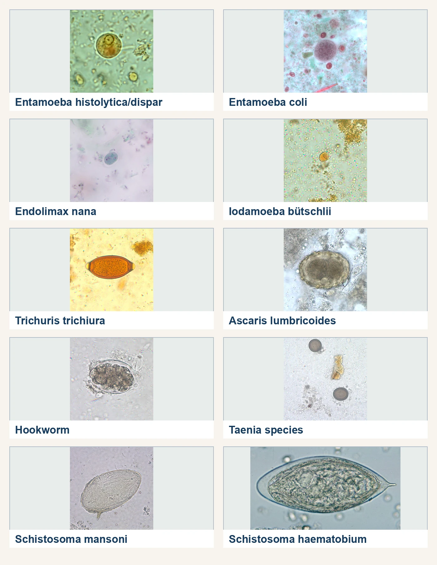
| E. histolytica/E. dispar/E. moshkovskii/E. bangladeshi complex | 10 to 60 µm, usually 15 to 20; one nucleus with a small usually central karyosome and fine, even peripheral chromatin; directional motility may occur | 10 to 20 µm, usually 12 to 15; mature cyst has 4 nuclei; blunt-ended chromatoid bodies may be present | Morphology does not resolve the species. Ingested erythrocytes favor E. histolytica but are not species-definitive. |
| Entamoeba hartmanni | 5 to 12 µm; resembles the E. histolytica complex | 5 to 10 µm; 4 nuclei when mature | Small size supports identification. Fixation shrinkage and natural size overlap occur, so measure several organisms. |
| Entamoeba coli | 15 to 50 µm; coarse, irregular peripheral chromatin and a large often eccentric karyosome | 10 to 35 µm; usually 8 nuclei when mature, occasionally more; pointed or splinterlike chromatoid bodies | The mature 8-nucleus cyst is the strongest distinction from the 4-nucleus complex. |
| Entamoeba polecki | 10 to 25 µm; one nucleus | 9 to 18 µm; usually 1 nucleus, rarely 2; variable pointed chromatoid bodies and sometimes an inclusion mass | A mononucleate cyst can resemble an immature cyst of another Entamoeba. |
| Endolimax nana | 6 to 12 µm; large blotlike karyosome and no peripheral chromatin | 5 to 10 µm; 4 nuclei when mature | Large irregular karyosomes, absent peripheral chromatin, and small size. |
| Iodamoeba buetschlii | 8 to 20 µm; large karyosome surrounded by achromatic granules; no peripheral chromatin | 5 to 20 µm; 1 nucleus and a compact iodine-staining glycogen mass | A single nucleus plus the sharply defined glycogen vacuole. |
| Dientamoeba fragilis | 5 to 15 µm; usually 2 nuclei, sometimes 1; each karyosome commonly fragments into 4 to 8 granules; angular or broad pseudopods | Rare putative cyst and precyst forms have been described, but their role is unresolved | Prompt fixation and permanent stain or NAAT. Concentrated wet mounts commonly miss it. |
Only E. histolytica is an established invasive member of its morphologic complex. E. dispar, E. moshkovskii, and E. bangladeshi can be indistinguishable from it by light microscopy. A validated E. histolytica-specific antigen assay or NAAT is needed for a species-level result. Molecular methods may also resolve the other members. Serology is most useful for extraintestinal amebiasis, especially amebic liver abscess, but antibody may persist after infection and can reflect prior exposure. Stool microscopy has low sensitivity in isolated liver abscess.8
The complete life cycle and pathogenic significance of D. fragilis remain unsettled. Human stool usually contains trophozoites, asymptomatic detection is common, and transmission within Enterobius eggs remains a hypothesis.
Blastocystis sp. is a stramenopile (a eukaryote in a lineage separate from the protozoa and fungi) retained in diagnostic parasitology. The usual fecal form is a spherical or oval vacuolar form, 5 to 40 µm and commonly 8 to 10 µm, with a large central body and a thin rim of cytoplasm containing peripheral nuclei. On trichrome the central body stains variably from red to blue and the nuclei stain purple. The proposed cyst and transmission cycle are not fully established. Because the organism occurs in symptomatic and asymptomatic people, detection alone does not prove that it caused the patient's diarrhea.9
Flagellates, the human ciliate, and urogenital trichomoniasis
Scroll sideways to see all columns
| Organism and stage | Size and morphology | Infective and diagnostic stages |
|---|---|---|
| Giardia duodenalis trophozoite | 10 to 20 µm; pear-shaped and bilaterally symmetric, with 2 nuclei, a ventral adhesive disk, paired median bodies, axonemes, and 8 flagella; falling-leaf motility | Trophozoites are diagnostic, especially in loose stool and duodenal material. |
| G. duodenalis cyst | 8 to 19 µm, usually 10 to 14; oval, with 2 nuclei when immature and 4 when mature; axonemes and median bodies lie within the cyst | The mature cyst is infective and immediately infectious when passed; each cyst releases 2 trophozoites after excystation. Humans are definitive hosts and no intermediate host is required. |
| Chilomastix mesnili | Trophozoite 6 to 24 µm with stiff rotary motility, a cytostome, and a spiral groove; cyst 6 to 10 µm, lemon-shaped with a hyaline nipple | The cyst is infective. Cysts and trophozoites are diagnostic. This is a nonpathogenic commensal. |
| Pentatrichomonas hominis | Trophozoite 6 to 20 µm with rapid jerky motility and an undulating membrane along the body; no recognized cyst | The trophozoite is the recognized stage. This is a nonpathogenic commensal. |
| Enteromonas hominis | Trophozoite 4 to 10 µm; cyst 4 to 10 µm, oval or elongated with 1 to 4 nuclei, often 2 at opposite ends | Cyst is infective. Both stages are diagnostic. Nonpathogenic. |
| Retortamonas intestinalis | Trophozoite 4 to 9 µm with a prominent cytostome; cyst 4 to 9 µm, pear- or lemon-shaped with 1 nucleus | Cyst is infective. Both stages are diagnostic. Nonpathogenic. |
| Balantioides coli (also reported as Balantidium coli or Neobalantidium coli) | Trophozoite 40 to 200 µm, covered in cilia, with rotary or boring motility and a bean-shaped macronucleus; cyst 50 to 70 µm | The cyst is infective. Cysts and trophozoites are diagnostic. Pigs are the principal reservoir; infection follows ingestion of fecally contaminated food or water. |
Stool antigen, direct fluorescent antibody, or NAAT usually detects Giardia more sensitively than a single O&P examination. Duodenal aspirate, string-test material, or small-bowel biopsy may show trophozoites when stool tests remain negative.10
Trichomonas vaginalis is a urogenital trichomonad with no recognized cyst. The 7 to 30 µm trophozoite has 4 anterior flagella, a recurrent flagellum associated with an undulating membrane extending about half the body, one anterior nucleus, and a posteriorly projecting axostyle. NAAT is preferred because wet-mount sensitivity is only 44% to 68% and falls rapidly when examination is delayed. Specimen types cleared by the Food and Drug Administration (FDA) differ by platform, and some assays accept male urine as well as female genital or urine specimens. A Papanicolaou test suggesting T. vaginalis requires confirmation with a sensitive diagnostic test.11
Coccidia and microsporidia
Scroll sideways to see all columns
| Organism | Fecal morphology and stage | Life cycle and laboratory detection |
|---|---|---|
| Cryptosporidium spp. | Round 3 to 6 µm oocyst, usually 4 to 5, containing 4 naked sporozoites and no sporocysts | Thick-walled sporulated oocysts are shed and immediately infectious. Thin-walled oocysts are involved in autoinfection. Modified acid-fast staining is less sensitive than DFA, antigen testing, or NAAT. Routine methods do not determine species. |
| Cyclospora cayetanensis | Spherical 7.5 to 10 µm unsporulated oocyst; variably acid-fast and often seen as an unstained wrinkled ghost | Environmental sporulation takes days to weeks and produces 2 sporocysts with 2 sporozoites each. Freshly passed oocysts are not infectious, so direct transmission is unlikely. Oocysts autofluoresce blue or green under the appropriate excitation. Hot safranin stains them more uniformly. |
| Cystoisospora belli | Ellipsoidal, usually 25 to 30 by 10 to 19 µm; the usual fecal oocyst is immature with 1 sporoblast; variably acid-fast | Environmental maturation produces 2 sporocysts with 4 sporozoites each. The oocyst wall autofluoresces. Repeated concentrated stools, duodenal material, or small-bowel biopsy may be needed. |
| Sarcocystis hominis and S. suihominis | Intact sporulated oocyst about 15 to 20 µm; the fragile wall often ruptures, leaving free sporocysts. S. hominis sporocysts are 13 to 17 µm; S. suihominis about 11 to 15 µm | Humans are definitive hosts after ingesting bradyzoite-containing sarcocysts in beef or pork. Cattle and pigs are the respective intermediate hosts. Sporulated oocysts or sporocysts are diagnostic and autofluoresce. Morphology does not separate the two species reliably. |
Cryptosporidium occupies an intracellular but extracytoplasmic position at the intestinal brush border. Because the oocyst is immediately infectious and resists routine chlorination, person-to-person and waterborne transmission occur. Yeast can mimic the small oocysts by size, so confirm the identification with the acid-fast reaction, DFA, antigen testing, or NAAT. In severe immunodeficiency, infection may extend to the biliary, pancreatic, or respiratory tract.12
Cyclospora oocysts are about twice the diameter of Cryptosporidium oocysts and autofluoresce. Cryptosporidium oocysts do not autofluoresce. Shedding is low-level and intermittent, so concentrate and examine specimens collected on different days. No animal reservoir for C. cayetanensis has been confirmed. Cystoisospora shedding may also be scant despite profuse diarrhea, and peripheral eosinophilia may occur. Most routine trichrome stains do not identify either organism reliably.13,14
Microsporidia are obligate intracellular, spore-forming eukaryotes closely related to fungi. The resistant spore is infective and contains a coiled polar tubule that everts to inject the sporoplasm into a host cell. Enterocytozoon bieneusi spores measure about 0.8 to 1.4 µm. Encephalitozoon spp. and several other human microsporidia measure about 1.5 to 4 µm. Modified trichrome or Chromotrope 2R stains spores pink-red, often with an equatorial band. Quick-hot Gram-chromotrope stains them dark violet. Examination requires oil immersion and an experienced reader. Light microscopy does not reliably determine species. Polymerase chain reaction (PCR), species-specific antibodies, or ultrastructural examination provides species identification. Stool is used for enteric disease, and urine may contain spores during disseminated Encephalitozoon infection.15
Intestinal nematodes
Scroll sideways to see all columns
| Organism | Diagnostic stage and morphology | Infective stage, hosts, and important exceptions |
|---|---|---|
| Enterobius vermicularis | Egg 50 to 60 by 20 to 30 µm, colorless and flattened on one side; recover by morning perianal tape or adhesive paddle before defecation, washing, or dressing | Partially embryonated eggs deposited on the perianum become infective in 4 to 6 hours. Humans are the usual host; there is no intermediate host. Three consecutive morning collections improve recovery. |
| Trichuris trichiura | Unembryonated egg 50 to 55 by 20 to 25 µm, barrel- or lemon-shaped with a thick shell and prominent bipolar plugs | Eggs embryonate in soil in about 15 to 30 days and become infective when ingested. Humans are definitive hosts; there is no intermediate host. |
| Ascaris spp. | Fertile egg about 45 to 75 by 35 to 50 µm, thick-shelled and usually mammillated; unfertilized eggs may reach 90 µm and cannot become infective | Larvae develop inside fertile eggs after 18 days to several weeks. Ingested eggs hatch, and larvae migrate through liver and lungs before adults mature in the small intestine. Pig-derived Ascaris, often called A. suum, can infect humans. |
| Intestinal hookworms: Necator americanus, Ancylostoma duodenale, A. ceylanicum | Egg 60 to 75 by 35 to 40 µm, thin-shelled and usually segmented; species eggs are indistinguishable | Filariform L3 usually penetrate skin. A. duodenale and A. ceylanicum can also be acquired orally. Larvae migrate through lungs before adults attach to small-intestinal mucosa. Chronic heavy infection can cause iron-deficiency anemia and protein loss. |
| Trichostrongylus spp. | Egg 75 to 95 by 40 to 50 µm, thin-shelled and more elongated than hookworm, often with one tapered end; overlap can prevent a confident distinction | Filariform L3 develop on soil or vegetation and are ingested. Herbivorous mammals are usual definitive hosts. Humans are incidental definitive hosts. |
| Strongyloides stercoralis | Rhabditiform L1, 180 to 380 µm, with a short buccal canal and prominent genital primordium; eggs hatch in the mucosa and are rarely seen in ordinary stool | Filariform L3 penetrate skin. Parasitic females reproduce in the small intestine; larvae may enter a free-living cycle or cause internal and external autoinfection. Filariform larvae have a long esophagus and notched tail. |
| Capillaria (=Paracapillaria) philippinensis | Egg 35 to 45 by 20 to 25 µm, with a striated shell and inconspicuous flattened polar prominences; eggs, larvae, and adults may occur in stool | Humans acquire larvae in raw or undercooked freshwater fish and appear to be principal definitive hosts. Thin-shelled eggs can hatch in the intestine and cause autoinfection. |
Hookworm rhabditiform larvae have a long buccal canal and inconspicuous genital primordium, the reverse of Strongyloides. Hookworm larvae in stool usually indicate delayed processing or culture. Strongyloides larvae are passed from an active infection. Decorticated Ascaris eggs retain a thick smooth shell. Do not mistake them for hookworm eggs, which have a thin shell and segmented contents. The morphology and life cycles in this table follow current DPDx organism descriptions.6,16-20
Routine O&P microscopy has low sensitivity for uncomplicated strongyloidiasis. Serial fresh stools examined by Baermann sedimentation or Koga agar-plate culture improve larval recovery. Charcoal and Harada-Mori cultures are alternatives. Formalin-ethyl acetate processing can remove larvae. Agar culture plates can produce infective filariform larvae and must remain sealed and be handled with exposure precautions. Serology helps with screening and low-burden infection, but cross-reactions occur, a positive result does not distinguish active from past infection, and immunosuppression can produce false-negative results. PCR requires a validated non-formalin-fixed specimen.21
Autoinfection allows S. stercoralis to persist for decades. Hyperinfection is accelerated autoinfection with increased larvae along the usual gastrointestinal and pulmonary route. In dissemination, larvae spread to organs outside that route. Corticosteroids are the most important precipitant. Human T-cell lymphotropic virus type 1 (HTLV-1) infection, hematologic malignancy, transplantation, and other immunosuppression also increase risk. Larvae may appear in stool, sputum, or bronchoalveolar lavage. In severe disease, enteric bacteria can enter the blood or cerebrospinal fluid (CSF). Eosinophilia may be absent.21
Cestodes
Scroll sideways to see all columns
| Organism | Diagnostic morphology | Infective stage and host relationship |
|---|---|---|
| Taenia saginata, T. solium, T. asiatica | Egg about 30 to 40 µm with a thick radially striated embryophore and 6-hooked oncosphere; species eggs are indistinguishable. Gravid T. saginata proglottids usually have 12 to 30 primary uterine branches per side. T. solium has 7 to 13. | A cysticercus in beef, pork, or pig viscera infects the human definitive host and causes taeniasis. Cattle are intermediate hosts for T. saginata and pigs for T. solium and T. asiatica. Ingestion of T. solium eggs makes a human an accidental intermediate host and causes cysticercosis. |
| Hymenolepis nana | Egg 30 to 50 µm, colorless, with a 6-hooked oncosphere, polar thickenings, and 4 to 8 polar filaments | Eggs are immediately infective. Humans can serve as both definitive and intermediate hosts because the cysticercoid develops in an intestinal villus. Internal autoinfection occurs; arthropods are optional intermediate hosts. |
| Hymenolepis diminuta | Egg 70 to 85 by 60 to 80 µm, yellow-brown, without polar filaments | A cysticercoid in a grain beetle or other arthropod infects rodents, the usual definitive hosts. Humans are accidental definitive hosts. |
| Diphyllobothriids, including Dibothriocephalus and Adenocephalus | Unembryonated egg 55 to 75 by 40 to 50 µm with an often inconspicuous operculum and a small abopercular knob; eggs usually support family-level identification only | A copepod is the first intermediate host and fish are the second. The plerocercoid in fish infects humans and other fish-eating definitive hosts. Molecular testing is usually needed for species identification. |
| Dipylidium caninum | Eggs average 35 to 40 µm and usually occur in packets containing 5 to 15 or more; proglottids have 2 lateral genital pores | The cysticercoid in an adult dog or cat flea infects the definitive host. Humans, usually children, are accidental definitive hosts after swallowing a flea. |
Handle unfixed Taenia specimens as though they may contain immediately infectious T. solium eggs until the species is established. Undercooked pork containing cysticerci causes intestinal taeniasis. Cysticercosis follows ingestion of eggs shed by a human carrier. Egg microscopy cannot separate the human Taenia species or distinguish them from other taeniid eggs such as Echinococcus.22
H. nana can persist through internal autoinfection. Diphyllobothriid taxonomy has changed substantially, and many historical cases assigned to Dibothriocephalus latus lacked species confirmation. A minority of broad fish tapeworm infections cause vitamin B12 deficiency. Dipylidium caninum is usually recognized when motile proglottids or egg packets are recovered, and flotation may miss the heavy packets.23-25
Trematodes
Trematodes use a snail as the first intermediate host. Most infect humans through ingestion of a metacercaria in vegetation, fish, or crustaceans. Schistosome cercariae penetrate skin, and schistosomes have no second intermediate host. Most trematode eggs are operculated. Schistosome eggs are nonoperculated and contain a mature miracidium when passed.6
Scroll sideways to see all columns
| Organism | Diagnostic stage and specimen | Infective stage and hosts |
|---|---|---|
| Fasciola hepatica, F. gigantica, Fasciolopsis buski | Large operculated unembryonated eggs, about 130 to 150 by 60 to 90 µm, overlap and may require a group-level report. Fasciola eggs occur in stool, duodenal fluid, or bile during chronic biliary infection. Fasciolopsis adults and eggs are intestinal. | Metacercariae on aquatic vegetation infect humans. Herbivores are usual Fasciola definitive hosts. Humans and pigs are definitive hosts for Fasciolopsis. Acute fascioliasis may be egg-negative because egg production begins months after exposure. |
| Clonorchis sinensis and Opisthorchis spp. | Small embryonated eggs, about 19 to 35 by 10 to 20 µm, with a shouldered operculum and small abopercular knob; species are practically indistinguishable by egg morphology | Metacercariae in freshwater fish infect humans and other fish-eating definitive hosts after sequential development in snails and fish. Adults inhabit bile ducts. |
| Heterophyes heterophyes and Metagonimus yokogawai | Small embryonated operculated eggs in stool resemble opisthorchiid eggs and are not reliably separated from one another | Metacercariae in fish infect human and other fish-eating definitive hosts. Adults inhabit the small intestine. |
| Nanophyetus salmincola | Operculated eggs about 64 to 97 by 34 to 55 µm in stool | Metacercariae in salmonid fish infect humans and other fish-eating mammals. |
| Paragonimus spp. | Thick-shelled, operculated, unembryonated eggs about 68 to 118 by 39 to 67 µm; sputum is characteristic, and swallowed eggs may appear in stool | Metacercariae in freshwater crab or crayfish infect humans and other definitive hosts after development in snails and crustaceans. Serology helps when eggs are not recovered or disease is ectopic. |
| Schistosoma mansoni | Egg 114 to 180 by 45 to 70 µm with a prominent lateral spine; stool | Cercariae from freshwater snails penetrate skin; humans are definitive hosts. |
| S. japonicum and S. mekongi | S. japonicum egg 70 to 100 by 55 to 64 µm; S. mekongi about 50 to 80 by 40 to 65 µm; both have a small, inconspicuous lateral spine and occur in stool | Cercariae penetrate skin. Mammalian reservoir hosts are important for S. japonicum, and dogs can harbor S. mekongi. |
| S. haematobium | Egg 110 to 170 by 40 to 70 µm with a terminal spine; primarily urine | Cercariae penetrate skin; humans are definitive hosts. |
| S. intercalatum and S. guineensis | Elongated terminal-spined eggs, often 140 to 240 µm long; stool | Cercariae penetrate skin; humans are principal definitive hosts. |
Recent consumption of animal liver can explain Fasciola-compatible eggs in stool without an established infection. This dietary passage is called pseudofascioliasis. Record the observed eggs, review the collection history, and recommend repeat stool examination after several days without liver before interpreting the finding as infection. Egg morphology alone cannot establish whether the eggs originated in the patient.26
For suspected intestinal schistosomiasis, examine repeated stool specimens by a sedimentation or validated quantitative method. For S. haematobium, urine collected around noon to 3 PM and examined as centrifuged sediment or by standardized membrane filtration improves recovery. When African exposure could involve both intestinal and urinary species, examine both stool and urine. Serology supports diagnosis in previously untreated travelers or immigrants when eggs are not recovered, but antibody does not distinguish active from treated infection. Cercarial dermatitis, acute Katayama syndrome (the systemic illness that follows heavy cercarial exposure), and chronic egg-induced fibrosis are possible manifestations, and a patient may have any one of them without the others.26-29
Tissue helminths
Filariae
Humans are definitive hosts for the major filariae. An arthropod is the biological intermediate host and vector. Thick and thin stained blood films show morphology. Knott concentration or membrane filtration improves sensitivity. Match blood collection to periodicity unless an antigen or molecular method makes timing unnecessary.30,31
Scroll sideways to see all columns
| Organism | Diagnostic specimen and morphology | Timing and vector |
|---|---|---|
| Wuchereria bancrofti | Sheathed microfilaria, 240 to 300 µm, smoothly curved with a pointed tail whose tip lacks nuclei; blood | Usually nocturnal, with geographic subperiodic exceptions; mosquitoes. Circulating antigen testing applies to W. bancrofti and can be performed at any time. |
| Brugia malayi | Sheathed microfilaria, 175 to 230 µm, with separated subterminal and terminal tail nuclei; blood | Usually nocturnal or subperiodic; principally Mansonia and Aedes mosquitoes. Sheath staining is method-dependent. |
| Brugia timori | Sheathed microfilaria about 310 µm with a long cephalic space; blood | Nocturnal; Anopheles mosquitoes in parts of Indonesia. |
| Loa loa | Sheathed microfilaria, 230 to 250 µm, with nuclei extending to the tail tip; blood | Diurnal, best collected about 10 AM to 2 PM; Chrysops flies. |
| Onchocerca volvulus | Unsheathed dermal microfilaria, about 220 to 360 µm, with no nuclei in the tail tip; thin bloodless skin snip in saline; adults may occur in nodules | Nonperiodic; Simulium black flies. Microfilariae usually emerge from the skin snip within 30 to 60 minutes. |
| Mansonella perstans | Unsheathed blood microfilaria about 190 to 200 µm with a blunt tail and nuclei to the tip | Generally nonperiodic; Culicoides midges. |
| M. ozzardi | Unsheathed blood microfilaria about 160 to 205 µm with a pointed tail lacking terminal nuclei | Generally nonperiodic; biting midges or black flies depending on region. |
| M. streptocerca | Unsheathed dermal microfilaria about 180 to 240 µm with a hooked tail and nuclei to the tip; skin snip | Nonperiodic; Culicoides midges. |
Migrating larvae and tissue cestodes
Humans are accidental or dead-end hosts for many tissue helminths, so stool O&P is negative. Toxocara larvae do not mature into egg-producing adults in humans. Diagnosis is based on exposure, compatible visceral or ocular disease, and serology. Demonstration of larvae in tissue is definitive but uncommon. The same principle applies to Baylisascaris procyonis. Anisakis and Pseudoterranova larvae are usually recovered by endoscopy, surgery, or histology after raw marine fish or squid exposure. Cutaneous larva migrans is usually diagnosed clinically. Trichinella antibody may remain negative for 3 to 5 weeks after infection, so early negative serum may need a later specimen. Muscle biopsy confirms infection when larvae are seen, but it has limited sensitivity.32-35
In cysticercosis, a human becomes an intermediate host after ingesting T. solium eggs. Neurocysticercosis diagnosis combines exposure, clinical findings, both noncontrast computed tomography (CT) and magnetic resonance imaging (MRI), and a glycoprotein enzyme-linked immunotransfer blot when confirmation is needed. Antibody sensitivity is high with multiple viable cysts but poor with a single lesion or only calcifications. Antibody does not distinguish active from inactive infection.36
In echinococcosis, humans are accidental intermediate hosts. Canids are the definitive hosts of E. granulosus sensu lato, E. multilocularis, and E. vogeli. Wild felids are the definitive hosts of E. oligarthrus. Echinococcus granulosus sensu lato causes cystic echinococcosis, E. multilocularis causes infiltrative alveolar disease, E. vogeli causes polycystic neotropical disease, and E. oligarthrus causes a very rare unicystic form. Imaging is the main diagnostic basis and serology is supportive. Negative serology does not exclude disease, and invasive sampling requires a planned expert procedure because cyst leakage can cause an allergic reaction and secondary implantation.37
Humans acquire Dracunculus medinensis by drinking water containing infected copepods. Diagnosis is usually clinical when a gravid female emerges from a skin ulcer, and lesion fluid may contain larvae. Humans are accidental hosts for Angiostrongylus cantonensis, which causes eosinophilic meningitis after ingestion of larvae from mollusks, paratenic hosts, or contaminated produce. Human stool is not diagnostic. Specialized real-time PCR of cerebrospinal fluid is available through reference laboratories. A. costaricensis causes abdominal disease, and eggs, larvae, or adults may be found in intestinal surgical or biopsy tissue. They are not found in stool.38,39
Parasitic infection in immunocompromised and asplenic patients
Each type of immune defect predisposes to different parasitic infections. Advanced HIV and other disorders of cell-mediated immunity increase the likelihood that Cryptosporidium, Cystoisospora, and microsporidia will cause severe, prolonged, or relapsing diarrhea. Cyclospora can also produce prolonged disease. Primary antibody deficiencies, especially common variable immunodeficiency, are associated with persistent or recurrent giardiasis. Corticosteroid exposure is the strongest recognized trigger for Strongyloides hyperinfection. Asplenia or hyposplenia and impaired B-cell function, particularly B-cell lymphoma or rituximab exposure, increase the risk of severe or relapsing babesiosis.40-45
Scroll sideways to see all columns
| Organism | Important setting | Laboratory approach and limitations |
|---|---|---|
| Cryptosporidium spp. | Advanced HIV and other substantial immunosuppression can cause prolonged watery diarrhea, wasting, malabsorption, and biliary disease. | Test stool by DFA, modified acid-fast stain, antigen assay, or NAAT. DFA, antigen testing, and NAAT are more sensitive than acid-fast microscopy. Repeat sampling helps when shedding is low. Some rapid antigen tests produce false-positive results, and routine methods do not determine species. |
| Cyclospora cayetanensis | Illness can be severe, chronic, or relapsing with impaired cell-mediated immunity. Immunocompetent people also develop significant infection. | Request specific testing. Concentrate stool; identify 7.5 to 10 µm variably acid-fast, autofluorescent oocysts or use a validated NAAT. Panel coverage varies. |
| Cystoisospora belli | Advanced HIV and other immunocompromising conditions can cause profuse prolonged diarrhea, weight loss, and malabsorption. | Examine repeated concentrated stools by wet mount, modified acid-fast stain, or ultraviolet autofluorescence microscopy. NAAT availability varies. Duodenal material or small-bowel biopsy can establish infection when stool remains negative. |
| Microsporidia | E. bieneusi and Encephalitozoon intestinalis cause chronic diarrhea and malabsorption in advanced immunosuppression. Other species can cause ocular, urinary, respiratory, muscular, cerebral, or disseminated disease. | Routine O&P and routine trichrome are insufficient. Use a modified trichrome or fluorescent-brightener method at oil immersion, or PCR. Light microscopy rarely provides species identification. Examine urine when disseminated Encephalitozoon or related infection is suspected. |
| Strongyloides stercoralis | Corticosteroids, HTLV-1, hematologic malignancy, transplantation, and other immunosuppression can precipitate hyperinfection or dissemination. HIV infection alone has not been associated with the same increased risk. | Patients with compatible present or remote exposure are screened before major immunosuppression. Use serology with serial fresh-stool larval recovery or validated PCR. In hyperinfection, examine stool and respiratory specimens for larvae. A negative serology does not exclude infection in an immunosuppressed patient. |
| Giardia duodenalis | Persistent or recurrent disease is associated particularly with common variable immunodeficiency and other primary antibody deficiencies.45 | Use stool DFA, antigen testing, or NAAT. Collect specimens on different days when sensitivity remains a concern. Duodenal material may recover trophozoites after negative stool tests. |
| Toxoplasma gondii | Advanced HIV most often causes reactivation encephalitis. Transplant recipients may have reactivation, donor-derived infection, or disseminated disease. | Serum IgG identifies prior infection and risk but does not establish active disease. IgM and IgG avidity assess recent primary infection. They do not assess cerebral reactivation. CSF PCR is 96% to 100% specific but about 50% sensitive, and sensitivity falls after therapy begins. Tissue histology, immunohistochemistry, and PCR may be needed.46,47 |
| Babesia spp. | Asplenia or hyposplenia, B-cell lymphoma, rituximab, HIV, advanced age, and other immunosuppression increase the risk of severe or relapsing disease. | Confirm acute disease by manual examination of stained peripheral blood films or PCR. Multiple films may be needed at low parasitemia. Maltese-cross tetrads are diagnostic when present but uncommon. A single antibody result does not confirm acute infection because antibody can persist. |
The laboratory needs the immune defect and exposure history, because a routine O&P examination may omit acid-fast staining and microsporidia stains, and a multiplex panel may omit one or more opportunistic parasites. Persistent diarrhea with negative stool studies may require additional specimens, a different method, or examination of duodenal or biopsy material.41 At the bench, check which organisms the ordered test can detect before you report a negative result.
Practice
References
- Garcia LS, Arrowood M, Kokoskin E, et al. Practical guidance for clinical microbiology laboratories: laboratory diagnosis of parasites from the gastrointestinal tract. Clin Microbiol Rev. 2018;31(1):e00025-17. doi:10.1128/CMR.00025-17 Back to text
- Centers for Disease Control and Prevention. DPDx: Stool specimens, specimen collection. Accessed August 31, 2026. https://www.cdc.gov/dpdx/diagnosticprocedures/stool/specimencoll.html Back to text
- Centers for Disease Control and Prevention. DPDx: Stool specimens, specimen processing. Accessed August 31, 2026. https://www.cdc.gov/dpdx/diagnosticprocedures/stool/specimenproc.html Back to text
- Centers for Disease Control and Prevention. DPDx: Stool specimens, molecular diagnosis. Accessed August 31, 2026. https://www.cdc.gov/dpdx/diagnosticprocedures/stool/moleculardx.html Back to text
- Centers for Disease Control and Prevention. DPDx: Stool specimens, staining procedures. Accessed August 31, 2026. https://www.cdc.gov/dpdx/diagnosticprocedures/stool/staining.html Back to text
- Centers for Disease Control and Prevention. DPDx: Intestinal parasites, comparative morphology tables. Accessed August 31, 2026. https://www.cdc.gov/dpdx/diagnosticprocedures/stool/morphcomp.html Back to text
- Centers for Disease Control and Prevention. DPDx: Stool specimens, detection of parasite antigens. Accessed August 31, 2026. https://www.cdc.gov/dpdx/diagnosticprocedures/stool/antigendetection.html Back to text
- Centers for Disease Control and Prevention. DPDx: Amebiasis. Accessed August 31, 2026. https://www.cdc.gov/dpdx/amebiasis/index.html Back to text
- Centers for Disease Control and Prevention. DPDx: Blastocystis. Accessed August 31, 2026. https://www.cdc.gov/dpdx/blastocystis/index.html Back to text
- Centers for Disease Control and Prevention. DPDx: Giardiasis. Accessed August 31, 2026. https://www.cdc.gov/dpdx/giardiasis/index.html Back to text
- Centers for Disease Control and Prevention. Trichomoniasis. In: Sexually Transmitted Infections Treatment Guidelines, 2021. Accessed August 31, 2026. https://www.cdc.gov/std/treatment-guidelines/trichomoniasis.htm Back to text
- Centers for Disease Control and Prevention. DPDx: Cryptosporidiosis. Accessed August 31, 2026. https://www.cdc.gov/dpdx/cryptosporidiosis/index.html Back to text
- Centers for Disease Control and Prevention. DPDx: Cyclosporiasis. Accessed August 31, 2026. https://www.cdc.gov/dpdx/cyclosporiasis/index.html Back to text
- National Institutes of Health. Cystoisosporiasis: guidelines for the prevention and treatment of opportunistic infections in adults and adolescents with HIV. Updated April 23, 2025. Reviewed March 16, 2026. Accessed August 31, 2026. https://clinicalinfo.hiv.gov/en/guidelines/hiv-clinical-guidelines-adult-and-adolescent-opportunistic-infections/cystoisosporiasis Back to text
- Centers for Disease Control and Prevention. DPDx: Microsporidiosis. Accessed August 31, 2026. https://www.cdc.gov/dpdx/microsporidiosis/index.html Back to text
- Centers for Disease Control and Prevention. DPDx: Enterobiasis. Accessed August 31, 2026. https://www.cdc.gov/dpdx/enterobiasis/index.html Back to text
- Centers for Disease Control and Prevention. DPDx: Trichuriasis. Accessed August 31, 2026. https://www.cdc.gov/dpdx/trichuriasis/index.html Back to text
- Centers for Disease Control and Prevention. DPDx: Ascariasis. Accessed August 31, 2026. https://www.cdc.gov/dpdx/ascariasis/index.html Back to text
- Centers for Disease Control and Prevention. DPDx: Intestinal hookworm. Accessed August 31, 2026. https://www.cdc.gov/dpdx/hookworm/index.html Back to text
- Centers for Disease Control and Prevention. DPDx: Intestinal capillariasis. Accessed August 31, 2026. https://www.cdc.gov/dpdx/intestinalcapillariasis/index.html Back to text
- Centers for Disease Control and Prevention. DPDx: Strongyloidiasis. Accessed August 31, 2026. https://www.cdc.gov/dpdx/strongyloidiasis/index.html Back to text
- Centers for Disease Control and Prevention. DPDx: Taeniasis. Accessed August 31, 2026. https://www.cdc.gov/dpdx/taeniasis/index.html Back to text
- Centers for Disease Control and Prevention. DPDx: Hymenolepiasis. Accessed August 31, 2026. https://www.cdc.gov/dpdx/hymenolepiasis/index.html Back to text
- Centers for Disease Control and Prevention. DPDx: Diphyllobothriasis. Accessed August 31, 2026. https://www.cdc.gov/dpdx/diphyllobothriasis/index.html Back to text
- Centers for Disease Control and Prevention. DPDx: Dipylidium caninum. Accessed August 31, 2026. https://www.cdc.gov/dpdx/dipylidium/index.html Back to text
- Centers for Disease Control and Prevention. DPDx: Fascioliasis. Diagnostic findings. Accessed September 7, 2026. https://www.cdc.gov/dpdx/fascioliasis/index.html Back to text
- Centers for Disease Control and Prevention. DPDx: Clonorchiasis. Accessed August 31, 2026. https://www.cdc.gov/dpdx/clonorchiasis/index.html Back to text
- Centers for Disease Control and Prevention. DPDx: Paragonimiasis. Accessed August 31, 2026. https://www.cdc.gov/dpdx/paragonimiasis/index.html Back to text
- Centers for Disease Control and Prevention. DPDx: Schistosomiasis infection. Accessed August 31, 2026. https://www.cdc.gov/dpdx/schistosomiasis/index.html Back to text
- World Health Organization. Bench Aids for the Diagnosis of Filarial Infections. World Health Organization; 1997. Accessed August 31, 2026. https://www.cdc.gov/dpdx/resources/pdf/benchAids/Introduction_filariasis_who.pdf Back to text
- Centers for Disease Control and Prevention. DPDx: Onchocerciasis. Accessed August 31, 2026. https://www.cdc.gov/dpdx/onchocerciasis/index.html Back to text
- Centers for Disease Control and Prevention. DPDx: Toxocariasis. Accessed August 31, 2026. https://www.cdc.gov/dpdx/toxocariasis/index.html Back to text
- Centers for Disease Control and Prevention. DPDx: Baylisascariasis. Accessed August 31, 2026. https://www.cdc.gov/dpdx/baylisascariasis/index.html Back to text
- Centers for Disease Control and Prevention. DPDx: Anisakiasis. Accessed August 31, 2026. https://www.cdc.gov/dpdx/anisakiasis/index.html Back to text
- Centers for Disease Control and Prevention. DPDx: Trichinellosis. Accessed August 31, 2026. https://www.cdc.gov/dpdx/trichinellosis/index.html Back to text
- Centers for Disease Control and Prevention. DPDx: Cysticercosis. Accessed August 31, 2026. https://www.cdc.gov/dpdx/cysticercosis/index.html Back to text
- Centers for Disease Control and Prevention. DPDx: Echinococcosis. Accessed August 31, 2026. https://www.cdc.gov/dpdx/echinococcosis/index.html Back to text
- Centers for Disease Control and Prevention. DPDx: Dracunculiasis. Accessed August 31, 2026. https://www.cdc.gov/dpdx/dracunculiasis/index.html Back to text
- Centers for Disease Control and Prevention. DPDx: Angiostrongyliasis. Accessed August 31, 2026. https://www.cdc.gov/dpdx/angiostrongyliasis/index.html Back to text
- Centers for Disease Control and Prevention. Clinical care of strongyloides. Accessed August 31, 2026. https://www.cdc.gov/strongyloides/hcp/clinical-care/index.html Back to text
- Shane AL, Mody RK, Crump JA, et al. 2017 Infectious Diseases Society of America clinical practice guidelines for the diagnosis and management of infectious diarrhea. Clin Infect Dis. 2017;65(12):e45-e80. doi:10.1093/cid/cix669 Back to text
- National Institutes of Health. Cryptosporidiosis: guidelines for the prevention and treatment of opportunistic infections in adults and adolescents with HIV. Accessed August 31, 2026. https://clinicalinfo.hiv.gov/en/guidelines/hiv-clinical-guidelines-adult-and-adolescent-opportunistic-infections/cryptosporidiosis Back to text
- National Institutes of Health. Microsporidiosis: guidelines for the prevention and treatment of opportunistic infections in adults and adolescents with HIV. Updated November 14, 2023. Reviewed March 16, 2026. Accessed August 31, 2026. https://clinicalinfo.hiv.gov/en/guidelines/hiv-clinical-guidelines-adult-and-adolescent-opportunistic-infections/microsporidiosis Back to text
- Centers for Disease Control and Prevention. Clinical overview of babesiosis. Updated February 13, 2024. Accessed August 31, 2026. https://www.cdc.gov/babesiosis/hcp/clinical-overview/index.html Back to text
- Bonilla FA, Barlan I, Chapel H, et al. International consensus document (ICON): common variable immunodeficiency disorders. J Allergy Clin Immunol Pract. 2016;4(1):38-59. doi:10.1016/j.jaip.2015.07.025 Back to text
- National Institutes of Health. Toxoplasma gondii encephalitis: guidelines for the prevention and treatment of opportunistic infections in adults and adolescents with HIV. Accessed August 31, 2026. https://clinicalinfo.hiv.gov/en/guidelines/hiv-clinical-guidelines-adult-and-adolescent-opportunistic-infections/toxoplasmosis Back to text
- La Hoz RM, Morris MI. Tissue and blood protozoa including toxoplasmosis, Chagas disease, leishmaniasis, Babesia, Acanthamoeba, Balamuthia, and Naegleria in solid organ transplant recipients. Clin Transplant. 2019;33(9):e13546. doi:10.1111/ctr.13546 Back to text