Mycobacteria have a mycolic-acid-rich wall that resists acid-alcohol decolorization once dye has entered the cell. That property is acid-fastness, and it is why a dedicated acid-fast stain is the smear method when mycobacterial infection is suspected. The same wall makes cells stain poorly with crystal violet or safranin, so on a Gram stain they appear as faint, beaded, gram-neutral or ghost rods. Wright- and Papanicolaou-stained smears show similar ghost images.1
Acid-fast staining
Spread two to three drops of concentrated sediment thinly, air-dry, and heat-fix (15 minutes at 80 °C, or 1 to 2 hours at 65 to 70 °C on a slide warmer) before staining. Heat fixation does not reliably kill M. tuberculosis: tubercle bacilli have remained viable in flame-fixed and hot-plate-fixed smears in controlled studies, so handle unstained slides as potentially infectious and keep every smear preparation step inside a certified biological safety cabinet.2,3 The staining reagents, including phenol-containing carbol fuchsin and auramine mixtures, inactivate bacilli. For added safety, fix unstained smears with 5% phenol-ethanol inside the cabinet, which both fixes and inactivates them.3
Scroll sideways to see all columns
| Stain family | Members | How dye enters the wall | Reading magnification |
|---|---|---|---|
| Carbol fuchsin | Ziehl-Neelsen (heat), Kinyoun (cold; higher phenol replaces heat) | Heat or high phenol | 1,000× oil immersion |
| Fluorochrome | Auramine-O, auramine-rhodamine | Dye binds mycolic acid | 250× or 450× |
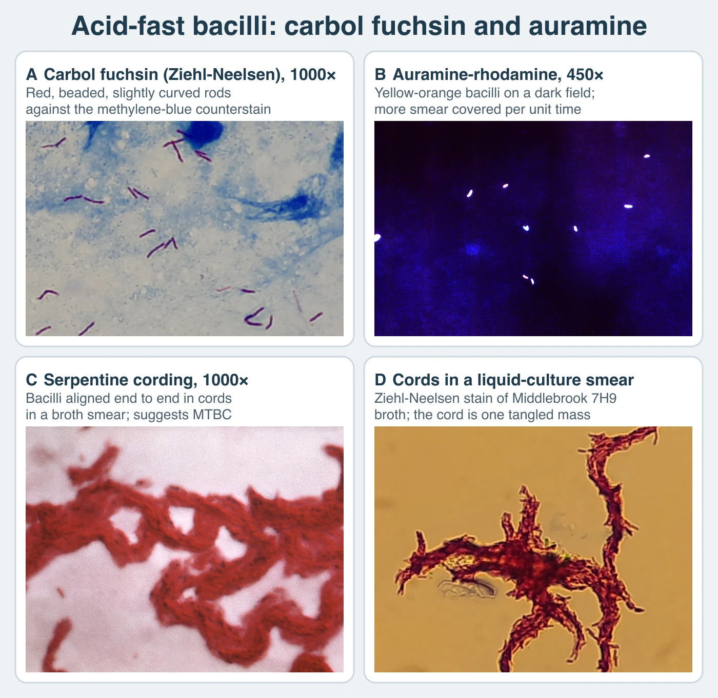
Fluorochrome screening covers far more of the smear per unit time than carbol fuchsin and is roughly 10% more sensitive than Ziehl-Neelsen against the same specimens. The Centers for Disease Control and Prevention (CDC) accepts either fluorochrome dye. Auramine-O and auramine-rhodamine perform equivalently, and the advantage comes from the fluorochrome method whichever dye is used. The World Health Organization (WHO) recommends light-emitting-diode fluorescence microscopy to replace mercury-vapor fluorescence microscopy and as a phased-in alternative to conventional Ziehl-Neelsen microscopy. That recommendation concerns the light source.1,4 Stained fluorochrome slides fade in light, so keep them covered and read them the same day.
Acid-fast bacilli (AFB) are red rods against the blue methylene-blue or green counterstain background with carbol fuchsin, and yellow-orange bacilli on a dark field with fluorochrome dyes. They are slightly curved rods, about 1 to 10 µm by 0.2 to 0.6 µm, often beaded or banded. Morphology does not identify species, but a few patterns are suggestive: serpentine cording in broth smears for M. tuberculosis complex (MTBC), driven by cord factor, trehalose 6,6'-dimycolate; long, broad, beaded, cross-barred bacilli, sometimes described as ladder-like, for M. kansasii; and short, pleomorphic, sometimes coccobacillary forms for M. avium complex (MAC). In tissue sections, macrophages packed with MAC are periodic acid-Schiff-positive and can mimic Whipple disease. An acid-fast stain distinguishes the two.1,5
Smear microscopy is highly specific, but a positive smear does not distinguish MTBC from nontuberculous mycobacteria or environmental acid-fast organisms. Sensitivity is about 20% to 80%, lowest in extrapulmonary and HIV-associated disease. According to CDC, roughly 5,000 to 10,000 bacilli per milliliter of specimen must be present for a positive smear. Culture can detect 10 to 100 organisms per milliliter.1,4 Causes of false-positive smears (positive smear, negative culture) include nonviable organisms shed after therapy, staining or carryover cross-contamination, environmental mycobacteria in reagents or rinse water, unfiltered carbol fuchsin precipitate, contaminated immersion oil, inadequate decolorization, and specimen mix-up. Investigate a smear-positive, culture-negative combination before you attribute it to nonviable bacilli after therapy.6
Sensitivity is higher with cavitary pulmonary disease, more specimens, and species such as M. tuberculosis or M. kansasii that shed more organisms than some nontuberculous mycobacteria (NTM).1
Smear results are reported semiquantitatively on the Kent and Kubica 1+ through 4+ scale, which CDC uses.1 Grade fluorochrome counts in the column for the magnification used:
Scroll sideways to see all columns
| Organisms seen (carbol fuchsin, 1,000×) | Organisms seen (fluorochrome, 250×) | Organisms seen (fluorochrome, 450×) | Report |
|---|---|---|---|
| None | None | None | No AFB seen |
| 1-2 per 300 fields | 1-2 per 30 fields | 1-2 per 70 fields | Report the exact count; request a repeat specimen |
| 1-9 per 100 fields | 1-9 per 10 fields | 2-18 per 50 fields | 1+ |
| 1-9 per 10 fields | 1-9 per field | 4-36 per 10 fields | 2+ |
| 1-9 per field | 10-90 per field | 4-36 per field | 3+ |
| >9 per field | >90 per field | >36 per field | 4+ |
A negative smear does not exclude tuberculosis. CDC expects an AFB smear result within 24 hours of specimen collection when the specimen reaches the laboratory promptly.1 Acid-fast organisms other than MTBC also stain, and rapidly growing mycobacteria can stain poorly or fail to fluoresce with fluorochrome dyes. When a rapid grower is likely, as in a postsurgical wound, restain a negative fluorochrome smear with carbol fuchsin before reporting it negative. M. fortuitum, M. abscessus, and M. chelonae are the species most often implicated.5
Mycobacterium tuberculosis complex
MTBC includes M. tuberculosis (the dominant human pathogen), M. bovis (cattle and other warm-blooded animals, including humans; the bacille Calmette-Guérin (BCG) vaccine strain is derived from it and can cause disseminated disease), M. africanum (West Africa), M. caprae (mainly goats and cattle), M. microti (voles and other small mammals), M. canettii, M. pinnipedii (pinnipeds), M. mungi, M. suricattae, and M. orygis, which is increasingly recognized in human disease.7,8 The proposal to split the genus into names such as Mycobacteroides and Mycolicibacterium has not been adopted by CDC, the Clinical and Laboratory Standards Institute (CLSI), or clinical laboratory practice, and United States laboratories report Mycobacterium names. Transmission is mainly inhalation of 1 to 5 µm droplet nuclei generated by coughing, shouting, or singing. Surface contact does not transmit MTBC. Direct inoculation of abraded skin is an occupational risk when infected tissue is handled. M. bovis also spreads through unpasteurized milk and from infected humans back to cattle via urine when genitourinary infection is present.1
Close contact with known or suspected active tuberculosis, birth in or travel to a high-prevalence country, congregate settings, health-care work with at-risk patients, and drug or alcohol use disorder increase the chance of acquiring infection. HIV, age under 5 years, immunosuppressive therapy including tumor necrosis factor-alpha antagonists, infection acquired in the past 2 years, prior inadequate treatment, silicosis, diabetes, chronic renal failure, certain cancers, gastrectomy or jejunoileal bypass, weight under 90% of ideal, and cigarette smoking increase the chance of progression to disease. About 5% to 10% of infected people progress to active tuberculosis over a lifetime, roughly half of them within the first 2 years after infection, and untreated HIV coinfection raises that risk sharply.1
Inhaled bacilli reach alveoli and are phagocytosed by macrophages that cannot kill them, because the bacilli block phagosome-lysosome fusion and survive intracellularly. Infected macrophages migrate to tracheobronchial nodes, present antigen, and seed the lung apices and distant sites (kidney, long-bone epiphyses, vertebral bodies, meninges). Cytokine-activated macrophages then drive caseation. A residual apical nidus can reactivate years later. The primary Ghon lesion is a 1 to 2 cm subpleural focus in the lower part of an upper lobe or the upper part of a lower lobe. Together with the draining-node lesion it is the Ghon complex.1
Pulmonary disease usually begins gradually, with cough lasting more than 2 weeks, fever, night sweats, weight loss, fatigue, hemoptysis, or pleuritic chest pain. Extrapulmonary disease concentrates in young children, older adults, and immunocompromised persons, especially with HIV coinfection.1
Nontuberculous mycobacteria
Runyon grouped NTM by pigment and growth rate on solid media: group I photochromogens pigment only after light exposure; group II scotochromogens pigment in dark or light; group III nonchromogens remain colorless; group IV rapid growers form visible colonies within 7 days on subculture. The scheme is obsolete for identification. Unrelated species share a Runyon phenotype, pigmentation varies with growth conditions, and liquid culture excludes light, so Runyon grouping cannot be done from a broth system. Current guidelines require species-level identification of clinically significant NTM by molecular methods or matrix-assisted laser desorption/ionization time-of-flight (MALDI-TOF) mass spectrometry. Runyon groups remain in use as descriptive terms. M. kansasii and M. marinum are group I photochromogens. M. gordonae, M. scrofulaceum, and M. szulgai are group II scotochromogens. MAC and M. xenopi are group III nonchromogens. M. abscessus, M. chelonae, and M. fortuitum are group IV rapid growers. More than 150 NTM species are now recognized.9
Scroll sideways to see all columns
| Organism / group | Disease pattern | Distinguishing notes |
|---|---|---|
| M. avium complex (MAC) | Most frequently isolated mycobacterium in US laboratories. Pulmonary disease mimicking tuberculosis (TB) or presenting as bronchiectasis; disseminated disease in advanced immunosuppression; childhood cervical lymphadenitis | Nonchromogen. Identify sterile-site isolates after cardiac surgery to species (M. chimaera and heater-cooler units) |
| M. genavense | Disseminated disease in HIV, resembling disseminated MAC | Requires mycobactin J and prolonged incubation |
| M. gordonae | Rarely pathogenic; common laboratory and tap-water contaminant | Slow scotochromogen; early growth can mimic MTBC; identify by DNA probe or sequencing |
| M. haemophilum | Cutaneous nodules or ulcers in immunocompromised hosts; childhood lymphadenitis | Requires hemin or ferric ammonium citrate; optimal growth 30-32 °C |
| M. kansasii | Chronic cavitary upper-lobe disease; extrapulmonary and disseminated disease | Long, broad, cross-barred bacilli; photochromogen |
| M. marinum | Skin granuloma after water exposure through broken skin | Fastest slow grower (~2 weeks); optimal growth 30-33 °C; photochromogen |
| M. ulcerans | Buruli ulcer, a progressive cutaneous ulcer caused by the mycolactone toxin | Optimal growth 30-33 °C; culture is slow and may need more than 8 weeks; IS2404 polymerase chain reaction (PCR) is the usual diagnostic target |
| M. xenopi | Pulmonary disease with preexisting lung disease or immunosuppression | "Bird's nest" colonies; classically a group III nonchromogen, though pale yellow pigment can develop as cultures age17; grows at 42 °C; hot-water-system pseudo-outbreaks |
| M. szulgai | Pulmonary and extrapulmonary disease similar to M. kansasii | Scotochromogenic at 37 °C, photochromogenic at 25 °C |
| M. scrofulaceum | Childhood cervical lymphadenitis, now uncommon | Group II scotochromogen |
| Rapid growers (M. fortuitum group; M. chelonae / M. abscessus complex) | Post-procedure skin and soft-tissue infection; chronic pulmonary disease in cystic fibrosis or bronchiectasis (usually M. abscessus); prosthetic-valve endocarditis; rare keratitis | Visible colonies within 7 days on subculture; primary isolation from a decontaminated specimen can take longer |
The 7-day rapid-grower criterion is defined on subculture to solid medium. After decontamination injury and with a low starting burden, a rapid grower can take weeks to appear on primary culture, so a slow primary isolation does not exclude a rapid grower.9
M. leprae has never been cultured in vitro. It is propagated in the mouse footpad over about 6 months and in the nine-banded armadillo, which is also a natural reservoir with documented zoonotic transmission in the southern United States. Its doubling time is about 12 to 14 days, among the slowest of known bacteria, and it grows best at cooler body sites such as skin and nasal mucosa. Transmission is principally person-to-person via nasal aerosols from lepromatous disease. Diagnosis uses skin-slit smear and lesion biopsy. In tissue the organism is demonstrated with the Fite-Faraco stain, because routine Ziehl-Neelsen preparation can strip its weakly acid-fast cells. Hansen disease is nationally notifiable in the United States. M. lepromatosis, the second species, is identified genomically and is associated with diffuse lepromatous leprosy mainly in Mexico.10
Disease versus colonization
The respiratory tract can be colonized or transiently contaminated with NTM without disease, so interpret a culture result against clinical and radiographic findings. The joint criteria of the American Thoracic Society, European Respiratory Society, European Society of Clinical Microbiology and Infectious Diseases, and Infectious Diseases Society of America (ATS/ERS/ESCMID/IDSA) for NTM pulmonary disease require compatible symptoms, compatible imaging (nodular or cavitary opacities, or multifocal bronchiectasis with small nodules on computed tomography), and a microbiologic standard: at least two separate expectorated sputum cultures positive for the same species, ideally collected at least 1 week apart; or one positive bronchial wash or bronchoalveolar lavage culture; or a lung biopsy with compatible histopathology and a positive culture. A single positive sputum culture therefore does not establish disease. NTM isolated from normally sterile sites, including blood, is always clinically significant.9
Specimen collection and decontamination
Scroll sideways to see all columns
| Disease pattern | Typically implicated organisms | Preferred specimen(s) |
|---|---|---|
| Pneumonia | MTBC, M. kansasii, MAC, M. abscessus | Early-morning sputum (3 consecutive days), bronchoalveolar lavage (BAL), lung tissue, pleural fluid, gastric contents (children who cannot produce sputum) |
| Disseminated disease | MTBC, MAC | Blood, bone marrow, involved tissue |
| Lymphadenitis | MTBC, MAC | Lymph-node aspirate or biopsy |
| Skin / soft tissue | Rapid growers, M. marinum, M. haemophilum, M. ulcerans | Aspirate or biopsy; swabs are unsuitable |
| Leprosy | M. leprae, M. lepromatosis | Skin-slit smear, lesion biopsy |
| Musculoskeletal | MTBC, rapid growers, M. marinum, M. kansasii | Joint fluid, synovium, bone |
| Meningitis / brain abscess | MTBC | Cerebrospinal fluid (CSF), abscess material |
| Genitourinary | M. tuberculosis, M. bovis BCG | Early-morning urine, involved tissue |
| Gastrointestinal / peritonitis | M. tuberculosis, M. bovis, MAC | Tissue, peritoneal fluid or biopsy |
| Hepatitis / pericarditis | MTBC, MAC | Liver tissue; pericardium or pericardial fluid |
| Catheter-related infection | Rapid growers | Blood |
Swabs are unacceptable for mycobacterial culture when fluid or tissue can be obtained, because they trap organisms in fibers, dry out, and provide too little material for digestion and decontamination. Specimens must never be submitted in formalin or any fixative, which kills organisms and is a rejection criterion for culture. Submit tissue fresh in a sterile container with a small amount of sterile saline. A separate formalin-fixed sample may accompany it for histology only. Blood for mycobacterial culture requires a dedicated method, a mycobacterium-specific bottle such as Myco/F Lytic or a lysis-centrifugation tube, held for up to 42 days. Sodium polyanetholesulfonate and heparin are acceptable anticoagulants. EDTA is inhibitory and unacceptable. Collect urine as a first-morning voided specimen. A 24-hour pooled collection is unacceptable because it dilutes the specimen and allows overgrowth. Stool culture for AFB is an ancillary test. When disseminated MAC is suspected, mycobacterial blood culture is the recommended method. A positive stool culture may reflect passage of organisms through the gut without disseminated disease.11,5
CDC asks for at least three consecutive sputum specimens, each collected 8 to 24 hours apart, with at least one early-morning specimen. The target volume is 5 to 10 mL, and pooled 24-hour sputum collections are unacceptable. Collect them in an airborne-infection isolation room or other well-ventilated space. Refrigerate specimens that cannot be processed promptly and process them within 24 hours, because mycobacteria lose viability and contaminants overgrow in delayed specimens. Sterile-site specimens may be inoculated directly. Specimens from sites with normal flora must be liquefied, decontaminated, and concentrated, or faster-growing flora will overgrow the mycobacteria. Perform all processing inside a certified Class II biological safety cabinet. MTBC specimen processing follows biosafety level 2 (BSL-2) practice. Manipulations of cultures and concentrated suspensions follow biosafety level 3 (BSL-3) practices and containment per Biosafety in Microbiological and Biomedical Laboratories (BMBL). Ventilation, respirator, and room requirements are covered in the biosafety section of the mycobacterial susceptibility lesson.1,2
The standard NALC-NaOH method uses an equal volume of freshly prepared digestant: equal parts 4% NaOH and 2.9% sodium citrate, with 0.5 g of NALC powder per 100 mL added immediately before use, used within 24 hours. NaOH is the decontaminant that kills faster-growing flora. NALC liquefies mucus so the digestant reaches organisms trapped in it. A shorter, gentler NaOH exposure is then sufficient. Sodium citrate binds heavy-metal ions in the specimen that would otherwise inactivate NALC. Vortex 15 to 30 seconds and let stand 15 minutes at room temperature. Do not exceed 15 minutes, because NaOH also kills some mycobacteria and longer contact kills more.12 Add sterile phosphate buffer pH 6.8 to about 45 to 50 mL to stop decontamination, then centrifuge at 3,000 to 3,900 × g for 15 to 20 minutes, refrigerated, in aerosol-tight sealed safety cups. Centrifuging a digest that may contain TB in unsealed cups is a recognized aerosol hazard. Decant and resuspend the sediment in 1.5 to 2 mL of sterile buffer. Never use non-sterile or deionized water, which introduces environmental mycobacteria such as M. gordonae.2,11
Specimens from cystic fibrosis or bronchiectasis patients overgrown with Pseudomonas receive a second decontamination with 5% oxalic acid after the NALC-NaOH step, because Pseudomonas survives standard NaOH exposure. Gastric aspirates must be processed within 4 hours or neutralized with sodium carbonate (about 100 mg per 5 to 10 mL), since gastric acid kills mycobacteria during any delay. Stool is diluted and filtered through gauze before decontamination. Urine sediments are concentrated by centrifugation at 3,000 to 3,600 × g for 30 minutes before processing.11,5
Contamination rate is the routine quality indicator for this bench. Some contamination is expected because the decontaminant must be mild enough for mycobacteria to survive it. The usual expected rate is about 2 to 5% of inoculated solid media and 7 to 8% of liquid media containing antimicrobials. A rate below the expected range suggests over-decontamination that kills mycobacteria along with contaminants, usually with a rise in smear-positive, culture-negative specimens. A rate above the range suggests under-decontamination, delayed transport, or contaminated reagents. Investigate and act on observed rates outside the expected range.11,16
Culture methods
Inoculate both broth and solid media. Broth detects growth sooner and in slightly higher overall yield. Solid media show colony morphology and pigmentation, provide isolated colonies for identification and susceptibility testing, and detect mixed cultures. In a large multicenter comparison, solid media detected roughly one in five positive cultures that liquid media missed. A broth-only approach lowers yield and is not endorsed for routine practice.11,5 Culture remains the reference method for laboratory confirmation of tuberculosis and is required for full susceptibility testing and genotyping even when a nucleic acid amplification test (NAAT) is positive.7
Solid media are egg-based (Löwenstein-Jensen) and agar-based (Middlebrook 7H10, 7H11, selective 7H11). Malachite green in Löwenstein-Jensen suppresses bacteria that survive decontamination, and mycobacteria tolerate it. Middlebrook 7H11 is 7H10 with casein hydrolysate, which supports fastidious and drug-damaged strains, including isoniazid-resistant isolates, that grow poorly on plain 7H10. Selective media add antibiotics for heavily contaminated specimens. Cutaneous specimens need hemin or ferric ammonium citrate for M. haemophilum and mycobactin J for M. genavense. Incubate at 35 to 37 °C in 5 to 10% CO2, keeping tubed media slanted with caps loosened for about the first week for gas exchange, then tightened to limit dehydration. Protect cultures from light throughout, because light degrades malachite green and induces pigment in photochromogens, which invalidates pigment-based observations. Skin, soft-tissue, joint, and bone specimens are also cultured in a second set at 30 °C (28 to 32 °C) to recover M. marinum, M. haemophilum, M. ulcerans, and some M. chelonae. These species grow preferentially or exclusively at temperatures below the routine 35 to 37 °C set, and 30 °C falls within every growth optimum listed in the tables above.11,5
Three types of liquid system are in current United States use. The BACTEC MGIT 960 uses tubes of modified Middlebrook 7H9 broth with a fluorogenic, oxygen-quenching sensor at the bottom. Growth consumes dissolved oxygen, quenching is relieved, and fluorescence rises. VersaTREK bottles are sealed with a headspace pressure sensor that registers gas consumption and production. BacT/ALERT MP bottles carry a colorimetric CO2 sensor at the bottle base that changes color as growth produces CO2. The radiometric BACTEC 460TB is discontinued. All liquid systems add an enrichment supplement (OADC, supplying oleic acid, albumin, dextrose, and catalase) and an antimicrobial mixture (PANTA) to suppress contaminant regrowth. Omitting the enrichment delays or prevents MTBC growth, and omitting the antimicrobials raises contamination rates sharply.11,5
Time to detection and the hold time before a negative report are separate values. MTBC in liquid culture is detected in about 7 to 21 days, faster from smear-positive than smear-negative specimens. On solid media it averages 3 to 6 weeks. A culture may be reported negative only after a full hold: a minimum of 42 days (6 weeks) for liquid systems and 8 weeks for solid media, with extended holds when M. ulcerans or M. genavense is suspected. Do not report a positive broth directly. Make an AFB smear of the broth, Gram stain turbid bottles as well, and subculture the broth to solid mycobacterial medium and to a blood agar purity plate. Growth on the purity plate usually represents a nonmycobacterial contaminant or a mixed culture. Rapidly growing mycobacteria also grow on blood agar, so identify the growth under the platform-specific procedure before you call it contamination. Treat a broth that signals positive with no AFB on smear as a probable instrument false signal or contaminant, and re-incubate it per the manufacturer's protocol. Mycobacterial growth is reported only after AFB are seen.11,5
Laboratory cross-contamination of cultures is uncommon but documented, accounting for a few percent of MTBC-positive cultures in genotyping studies. Clues include a single positive culture from one of several specimens, scant growth, an unusually long time to detection, and a smear-negative specimen positive by culture after being processed in the same batch as a strongly positive specimen. Investigation follows the CDC false-positive toolkit and uses genotyping, which is also the method CDC applies nationally to every culture-positive United States TB case to detect outbreaks, laboratory cross-contamination, and unsuspected transmission.6
Scroll sideways to see all columns
| Organism | Colony morphology | Pigment | Time to growth | Notes |
|---|---|---|---|---|
| M. tuberculosis | Rough | Nonchromogenic (buff) | 3-6 wk | Niacin- and nitrate-positive |
| M. bovis | Rough; thin/transparent on Löwenstein-Jensen | Nonchromogenic | 4-6 wk | Niacin- and nitrate-negative; pyrazinamide-resistant from loss of pyrazinamidase (pncA); the classic thiophene-2-carboxylic acid hydrazide susceptibility test separates it from M. tuberculosis |
| MAC | Smooth to large opaque, sometimes rough | Nonchromogenic | 3-6 wk | Some strains pigment late; growth at 42 °C is strain-variable and only a presumptive clue |
| M. kansasii | Rough, sometimes smooth, β-carotene crystals | Photochromogenic | 3-6 wk | Rare non/scotochromogenic strains |
| M. gordonae | Smooth | Scotochromogenic | 3-6 wk | Common contaminant |
| M. marinum | Wrinkled/shiny to smooth | Photochromogenic | 2 wk | Optimal growth 30-33 °C |
| M. xenopi | Smooth, filamentous ("bird's nest") | Nonchromogenic; pigment may develop with age | 3-6 wk | Grows at 42 °C |
| M. haemophilum | Rough, sometimes smooth | Nonchromogenic | 3-6 wk | Requires hemin or ferric ammonium citrate; optimal growth 30-32 °C |
| M. szulgai | Smooth or rough | Scotochromogenic (37 °C) / photochromogenic (25 °C) | 3-6 wk | - |
| M. abscessus complex | Smooth, sometimes rough | Nonchromogenic | 1 wk or less on subculture | Rapid grower |
| M. fortuitum group | Smooth with occasional filamentous projections at the periphery | Nonchromogenic | 1 wk or less on subculture | Rapid grower |
| M. chelonae | Smooth | Nonchromogenic | 1 wk or less on subculture | Rapid grower |
Identification methods
The classical biochemical battery (catalase at 68 °C, Tween 80 hydrolysis, arylsulfatase, iron uptake, tellurite, urease) has been superseded as a primary identification method. Niacin and nitrate are still used to presumptively differentiate M. tuberculosis from other MTBC members, notably M. bovis, and from NTM while molecular confirmation is pending. Current methods are faster and more specific.5
Direct-specimen NAAT gives the fastest laboratory result when pulmonary TB is suspected. CDC recommends NAAT on at least one respiratory specimen from each patient with symptoms of pulmonary TB when the diagnosis is being considered and the result would change case management or control activities.7 Real-time PCR assays such as Xpert MTB/RIF, the cartridge cleared by the Food and Drug Administration (FDA) and used in United States laboratories, detect MTBC and, in the same run, rpoB mutations associated with rifampin resistance, usually in under 2 hours. Transcription-mediated amplification of rRNA is the other cleared format, reporting positive, equivocal, or negative. The successor Xpert MTB/RIF Ultra is WHO-recommended and adds multicopy targets (IS6110 and IS1081), a lower limit of detection, better sensitivity in smear-negative and paucibacillary disease, and a trace result category, but it is not FDA-cleared for the United States market. NAAT sensitivity is higher on smear-positive than smear-negative sputum. No format differentiates individual MTBC species. NAAT detects DNA from nonviable organisms, so it cannot be used to monitor treatment response. NAAT does not replace smear, culture, growth-based susceptibility testing, or genotyping.13,14
Interpretation follows CDC categories. MTBC detected with rifampin resistance detected has a high probability of rifampin resistance, requires immediate notification of the clinician and the public health authority, and needs additional rapid confirmation, because silent or low-level rpoB mutations lower the positive predictive value in low-prevalence settings. MTBC detected with resistance not detected still needs growth-based first-line testing. An indeterminate rifampin result also needs growth-based testing. When a smear-positive specimen gives a negative NAAT, retest the specimen, including for inhibitors. When NAAT is negative but clinical suspicion persists, repeat testing on an additional specimen.7,13
Chemiluminescent DNA probes identify a species or complex within 1 to 2 hours once adequate growth is available. Known probe limitations moved laboratories toward MALDI-TOF and sequencing: occasional failure to identify MAC grown in liquid medium, false-positive MAC results from broth aliquots, and rare false-positive MTBC results. Hologic discontinued its AccuProbe culture-identification probes in the United States in December 2022, so isolates are now usually identified by MALDI-TOF or sequencing.5,18
MALDI-TOF requires extraction to disrupt the mycolic-acid-rich wall. Perform the extraction on live growth inside the containment laboratory. The extraction inactivates MTBC, but each laboratory must validate inactivation under its own conditions before material is handled outside containment. A dedicated mycobacterial reference library is required because routine bacterial databases lack validated mycobacterial spectra. VITEK MS includes mycobacteria in its FDA-cleared database. The Bruker mycobacteria library has not been FDA-cleared in the United States. Most protocols use solid-medium growth. MALDI-TOF cannot resolve individual MTBC species, cannot resolve M. abscessus subspecies, and cannot reliably separate M. chimaera from M. intracellulare. For this reason, sequencing was needed to recognize the heater-cooler-associated M. chimaera outbreak isolates.5
Subspecies identification of M. abscessus is clinically important. M. abscessus subsp. abscessus and subsp. bolletii typically carry a functional erm(41) gene that confers inducible macrolide resistance, although the gene in a T28C sequevar within subsp. abscessus is nonfunctional. Subsp. massiliense usually carries a nonfunctional gene and remains macrolide-susceptible. The subspecies predicts the usual pattern. The susceptibility lesson explains why the extended-incubation read is required to detect inducible resistance. Subspecies identification requires sequencing of targets such as rpoB, hsp65, or erm(41) itself.9
Line-probe reverse-hybridization assays hybridize labeled PCR product to immobilized probes. WHO-endorsed MTBDRplus and MTBDRsl detect first- and second-line drug resistance from colonies, positive broth, or AFB smear-positive specimens. Genus and species line-probe panels identify NTM. These assays are used mainly outside the United States, where they are part of national testing algorithms.
Sequencing has the highest identification accuracy among these methods. 16S rRNA is the usual first target, but it cannot resolve individual MTBC members, M. abscessus subspecies, or M. chimaera from M. intracellulare, so many species need additional loci (rpoB, secA1, hsp65, gyrB, or the internal transcribed spacer). CLSI M48 is the standard for sequencing-based identification of mycobacteria. Whole-genome sequencing adds resistance markers and is the basis of CDC's national genotyping of United States TB isolates and of the WHO catalog of MTBC mutations. Phenotypic susceptibility testing is still performed alongside it because uncataloged mutations are missed.9,15
Overall turnaround is longer than the assay run time. A cartridge or probe run takes about an hour or two, but identification from culture requires growth first. The expected sequence is a positive smear the day of receipt, a NAAT result within about a day of specimen receipt, a positive MTBC culture signal in 1 to 3 weeks, and identification and susceptibility testing following after that, with final negative cultures incubated the full 42 days (liquid) or 8 weeks (solid).1,11 Interpret every AFB smear result, positive or negative, with the specimen type and the stain and magnification used, and correlate it with the culture result when that is available.
Practice
References
- Centers for Disease Control and Prevention. Core Curriculum on Tuberculosis: What the Clinician Should Know. 7th ed. CDC; 2021. Fluorescent microscopy scale adapted from Kent PT, Kubica GP. Public Health Mycobacteriology: A Guide for the Level III Laboratory. CDC; 1985. Accessed August 31, 2026. https://www.cdc.gov/tb/media/Core_Curriculum_TB_eBook.pdf Back to text
- Clinical and Laboratory Standards Institute. M48, Laboratory Detection and Identification of Mycobacteria. 3rd ed. CLSI; 2025. Accessed August 31, 2026. https://clsi.org/shop/standards/m48/ Back to text
- Allen BW. Survival of tubercle bacilli in heat-fixed sputum smears. J Clin Pathol. 1981;34(7):719-722. Accessed August 31, 2026. https://pmc.ncbi.nlm.nih.gov/articles/PMC493800/ Back to text
- World Health Organization. Fluorescent light-emitting diode (LED) microscopy for diagnosis of tuberculosis: policy statement. WHO; 2011. Accessed August 31, 2026. https://www.ncbi.nlm.nih.gov/books/NBK131934/ Back to text
- Martin I, Pfyffer GE, Parrish N. Mycobacterium: general characteristics, laboratory detection, and staining procedures. In: Carroll KC, Pfaller MA, Landry ML, et al, eds. Manual of Clinical Microbiology. 12th ed. ASM Press; 2019:558-575. Back to text
- Centers for Disease Control and Prevention. False-positive tuberculosis laboratory results: investigation toolkit. Reviewed April 22, 2024. Accessed August 31, 2026. https://www.cdc.gov/tb/php/false-positive-investigation-toolkit/ Back to text
- Centers for Disease Control and Prevention. Clinical and laboratory diagnosis for tuberculosis. Updated January 31, 2025. Accessed August 31, 2026. https://www.cdc.gov/tb/hcp/testing-diagnosis/clinical-and-laboratory-diagnosis.html Back to text
- Centers for Disease Control and Prevention. Reported tuberculosis in the United States, 2022. Appendix B: tuberculosis surveillance case definition glossary. Accessed August 31, 2026. https://www.cdc.gov/tb/statistics/reports/2022/Appendix-B.html Back to text
- Daley CL, Iaccarino JM, Lange C, et al. Treatment of nontuberculous mycobacterial pulmonary disease: an official ATS/ERS/ESCMID/IDSA clinical practice guideline. Clin Infect Dis. 2020;71(4):e1-e36. Accessed August 31, 2026. https://pmc.ncbi.nlm.nih.gov/articles/PMC7768748/ Back to text
- Centers for Disease Control and Prevention. About leprosy (Hansen's disease). Updated December 5, 2025. Accessed September 24, 2026. https://www.cdc.gov/leprosy/about/index.html Back to text
- Association of Public Health Laboratories. Mycobacterium tuberculosis: Assessing Your Laboratory. APHL; April 2019. Accessed September 24, 2026. https://aphl.org/docs/default-source/technical/ID-2019Apr-TB-Toolkit.pdf Back to text
- Kent PT, Kubica GP. Public Health Mycobacteriology: A Guide for the Level III Laboratory. CDC; 1985. Back to text
- Centers for Disease Control and Prevention. Xpert MTB/RIF assay. Reviewed April 22, 2024. Accessed August 31, 2026. https://www.cdc.gov/tb/php/laboratory-information/xpert-mtb-rif-assay.html Back to text
- World Health Organization. The use of Xpert MTB/RIF Ultra and Xpert MTB/RIF assays for diagnosis of tuberculosis: report of a Technical Expert Consultation. WHO; 2017. Accessed August 31, 2026. https://www.who.int/publications/i/item/WHO-HTM-TB-2017.04 Back to text
- World Health Organization. Catalogue of mutations in Mycobacterium tuberculosis complex and their association with drug resistance. WHO; 2021. Accessed September 24, 2026. https://www.who.int/publications/i/item/9789240028173 Back to text
- Forbes BA, Hall GS, Miller MB, et al. Practical guidance for clinical microbiology laboratories: mycobacteria. Clin Microbiol Rev. 2018;31(2):e00038-17. doi:10.1128/CMR.00038-17 Back to text
- Tomioka H. Bacteriology of mycobacteria: taxonomic and morphological characteristics. Nihon Rinsho. 1999;56(12):3001-3007. Japanese. The review's abstract lists M. xenopi among the Runyon group III nonphotochromogens. Accessed September 11, 2026. https://pubmed.ncbi.nlm.nih.gov/9883600/ Back to text
- Armstrong DT, Pretty L, D'Agostino K, Redhead-Harper R, Parrish N. Diagnostic accuracy of the Abbott SD Bioline MPT64 antigen test for identification of MTB complex in a U.S. clinical mycobacteriology laboratory. Heliyon. 2024;10(9):e30501. doi:10.1016/j.heliyon.2024.e30501 Back to text